Morphological changes in blood in pneumonia in children, taking into account the impact of COVID-19
DOI:
https://doi.org/10.15587/2519-4798.2025.348501Keywords:
pneumonia, morphological changes in blood, COVID-19, hemomicrocirculatory system, SARS-CoV-2, hypoxia, lung diseaseAbstract
The study of morphological changes in blood in pneumonia in children, in particular those accompanied by coronavirus infection, is an important aspect for improving diagnostics and predicting the occurrence of complications arising from the action of oxidative stress.
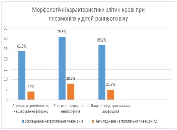
Objective: to identify and compare morphological changes in blood in children with pneumonia depending on the presence of COVID-19 infection, to find out which morphological indicators are associated with the severity of the disease and to assess the significance of morphological markers for the development of complications.
Materials and methods: A cohort study was conducted on the basis of the pulmonology department of the Ivano-Frankivsk Regional Children's Clinical Hospital of the Ivano-Frankivsk Regional Council in the period 2021-2025. Children aged 1-3 years with clinical and radiological confirmation of pneumonia were included. For this study, 60 young children (1-3 years old) diagnosed with community-acquired pneumonia were selected and divided into 2 groups according to the presence of complicated pneumonia.
Results: Using ultrastructural blood analysis, it was found that the most pronounced morphological changes are observed in complicated cases of COVID-19. Erythrocytes change their shape, some of them are destroyed, others participate in the formation of microthrombi. Among leukocytes, an increase in the number of segmented neutrophils, lymphocytes and monocytes is noted.
Conclusions. The results of the study will clarify the prognostic role of morphological changes in the blood in children with pneumonia, in particular with concomitant COVID-19 infection
References
- Horiuchi, Y., Hayashi, F., Iwasaki, Y., Matsuzaki, A., Nishibe, K., Kaniyu, K. et al. (2021). Peripheral granular lymphocytopenia and dysmorphic leukocytosis as simple prognostic markers in COVID‐19. International Journal of Laboratory Hematology, 43 (6), 1309–1318. https://doi.org/10.1111/ijlh.13696
- Sadigh, S., Massoth, L. R., Christensen, B. B., Stefely, J. A., Keefe, J., Sohani, A. R. (2020). Peripheral blood morphologic findings in patients with COVID‐19. International Journal of Laboratory Hematology, 42 (6). https://doi.org/10.1111/ijlh.13300
- Wang, F., Nie, J., Wang, H., Zhao, Q., Xiong, Y., Deng, L. et al. (2020). Characteristics of Peripheral Lymphocyte Subset Alteration in COVID-19 Pneumonia. The Journal of Infectious Diseases, 221 (11), 1762–1769. https://doi.org/10.1093/infdis/jiaa150
- Lippi, G., Plebani, M., Henry, B. M. (2020). Thrombocytopenia is associated with severe coronavirus disease 2019 (COVID-19) infections: A meta-analysis. Clinica Chimica Acta, 506, 145–148. https://doi.org/10.1016/j.cca.2020.03.022
- Mehta, P., McAuley, D. F., Brown, M., Sanchez, E., Tattersall, R. S., Manson, J. J. (2020). COVID-19: consider cytokine storm syndromes and immunosuppression. The Lancet, 395 (10229), 1033–1034. https://doi.org/10.1016/s0140-6736(20)30628-0
- Zini, G., Bellesi, S., Ramundo, F., d’Onofrio, G. (2020). Morphological anomalies of circulating blood cells in COVID‐19. American Journal of Hematology, 95 (7), 870–872. https://doi.org/10.1002/ajh.25824
- Merino, A., Vlagea, A., Molina, A., Egri, N., Laguna, J., Barrera, K. et al. (2020). Atypical lymphoid cells circulating in blood in COVID-19 infection: morphology, immunophenotype and prognosis value. Journal of Clinical Pathology, 75 (2), 104–111. https://doi.org/10.1136/jclinpath-2020-207087
- Tan, L., Wang, Q., Zhang, D., Ding, J., Huang, Q., Tang, Y.-Q. et al. (2020). Lymphopenia predicts disease severity of COVID-19: a descriptive and predictive study. Signal Transduction and Targeted Therapy, 5 (1). https://doi.org/10.1038/s41392-020-0148-4
- Yamada, T., Wakabayashi, M., Yamaji, T., Chopra, N., Mikami, T., Miyashita, H., Miyashita, S. (2020). Value of leukocytosis and elevated C-reactive protein in predicting severe coronavirus 2019 (COVID-19): A systematic review and meta-analysis. Clinica Chimica Acta, 509, 235–243. https://doi.org/10.1016/j.cca.2020.06.008
- Tuharov, Y., Dvorshchenko, K. (2024). Oxidative modification of proteins in blood plasma of patients with osteoarthritis after SARS-CoV2 Infection. Bulletin of Taras Shevchenko National University of Kyiv. Series: Biology, 97 (2), 22–27. https://doi.org/10.17721/1728.2748.2024.97.22-27
- Cecchini, R., Cecchini, A. L. (2020). SARS-CoV-2 infection pathogenesis is related to oxidative stress as a response to aggression. Medical Hypotheses, 143, 110102. https://doi.org/10.1016/j.mehy.2020.110102
- Forcados, G. E., Muhammad, A., Oladipo, O. O., Makama, S., Meseko, C. A. (2021). Metabolic Implications of Oxidative Stress and Inflammatory Process in SARS-CoV-2 Pathogenesis: Therapeutic Potential of Natural Antioxidants. Frontiers in Cellular and Infection Microbiology, 11. https://doi.org/10.3389/fcimb.2021.654813
- Georgieva, E., Ananiev, J., Yovchev, Y., Arabadzhiev, G., Abrashev, H., Abrasheva, D. et al. (2023). COVID-19 Complications: Oxidative Stress, Inflammation, and Mitochondrial and Endothelial Dysfunction. International Journal of Molecular Sciences, 24 (19), 14876. https://doi.org/10.3390/ijms241914876
- Spiezia, L., Boscolo, A., Poletto, F., Cerruti, L., Tiberio, I., Campello, E., Navalesi, P., Simioni, P. (2020). COVID-19-Related Severe Hypercoagulability in Patients Admitted to Intensive Care Unit for Acute Respiratory Failure. Thrombosis and Haemostasis, 120 (6), 998–1000. https://doi.org/10.1055/s-0040-1710018
- Tang, N., Li, D., Wang, X., Sun, Z. (2020). Abnormal coagulation parameters are associated with poor prognosis in patients with novel coronavirus pneumonia. Journal of Thrombosis and Haemostasis, 18 (4), 844–847. https://doi.org/10.1111/jth.14768
- Harris, C. K., Hung, Y. P., Nielsen, G. P., Stone, J. R., Ferry, J. A. (2021). Bone Marrow and Peripheral Blood Findings in Patients Infected by SARS-CoV-2. American Journal of Clinical Pathology, 155 (5), 627–637. https://doi.org/10.1093/ajcp/aqaa274
- Bahadur, S., Kalonia, T., Kamini, K., Gupta, B., Kalhan, S., Jain, M. (2021). Changes in peripheral blood in SARS CoV‐2 patients and its clinico‐pathological correlation: A prospective cross‐sectional study. International Journal of Laboratory Hematology, 43 (6), 1334–1340. https://doi.org/10.1111/ijlh.13720
- Cattaneo, M., Bertinato, E. M., Birocchi, S., Brizio, C., Malavolta, D., Manzoni, M. et al. (2020). Pulmonary Embolism or Pulmonary Thrombosis in COVID-19? Is the Recommendation to Use High-Dose Heparin for Thromboprophylaxis Justified? Thrombosis and Haemostasis, 120 (8), 1230–1232. https://doi.org/10.1055/s-0040-1712097
- Grobbelaar, L. M., Venter, C., Vlok, M., Ngoepe, M., Laubscher, G. J., Lourens, P. J. et al. (2021). SARS-CoV-2 spike protein S1 induces fibrin(ogen) resistant to fibrinolysis: implications for microclot formation in COVID-19. Bioscience Reports, 41 (8). https://doi.org/10.1042/bsr20210611
- Matharu, S. S., Nordmann, C. S., Ottman, K. R., Akkem, R., Palumbo, D., Cruz, D. R. D. et al. (2023). Deep learning, 3D ultrastructural analysis reveals quantitative differences in platelet and organelle packing in COVID-19/SARSCoV2 patient-derived platelets. Platelets, 34 (1). https://doi.org/10.1080/09537104.2023.2264978
Downloads
Published
How to Cite
Issue
Section
License
Copyright (c) 2025 Oksana Dutchuk, Zoryana Kocherha

This work is licensed under a Creative Commons Attribution 4.0 International License.
Our journal abides by the Creative Commons Attribution 4.0 International License copyright rights and permissions for open access journals.
Authors, who are published in this journal, agree to the following conditions:
1. The authors reserve the right to authorship of the work and pass the first publication right of this work to the journal under the terms of a Creative Commons Attribution 4.0 International License, which allows others to freely distribute the published research with the obligatory reference to the authors of the original work and the first publication of the work in this journal.
2. The authors have the right to conclude separate supplement agreements that relate to non-exclusive work distribution in the form in which it has been published by the journal (for example, to upload the work to the online storage of the journal or publish it as part of a monograph), provided that the reference to the first publication of the work in this journal is included.
3. Authors have the right to store the final accepted version of the article in an institutional, thematic, or any other repository to ensure visibility and accessibility.
