Eosinophilic sigmoiditis as a manifestation of severe gastrointestinal salmonellosis in children (clinical case)
DOI:
https://doi.org/10.15587/2519-4798.2025.348480Keywords:
children, salmonellosis, severe gastrointestinal forms, eosinophilic sigmoiditis, antibiotic resistanceAbstract
Salmonellosis in children represents a significant medical and social problem due to its high incidence and the emergence of multidrug-resistant Salmonella strains.
Objective. To analyze a clinical case of severe intestinal involvement in the gastrointestinal form of salmonellosis in a child.
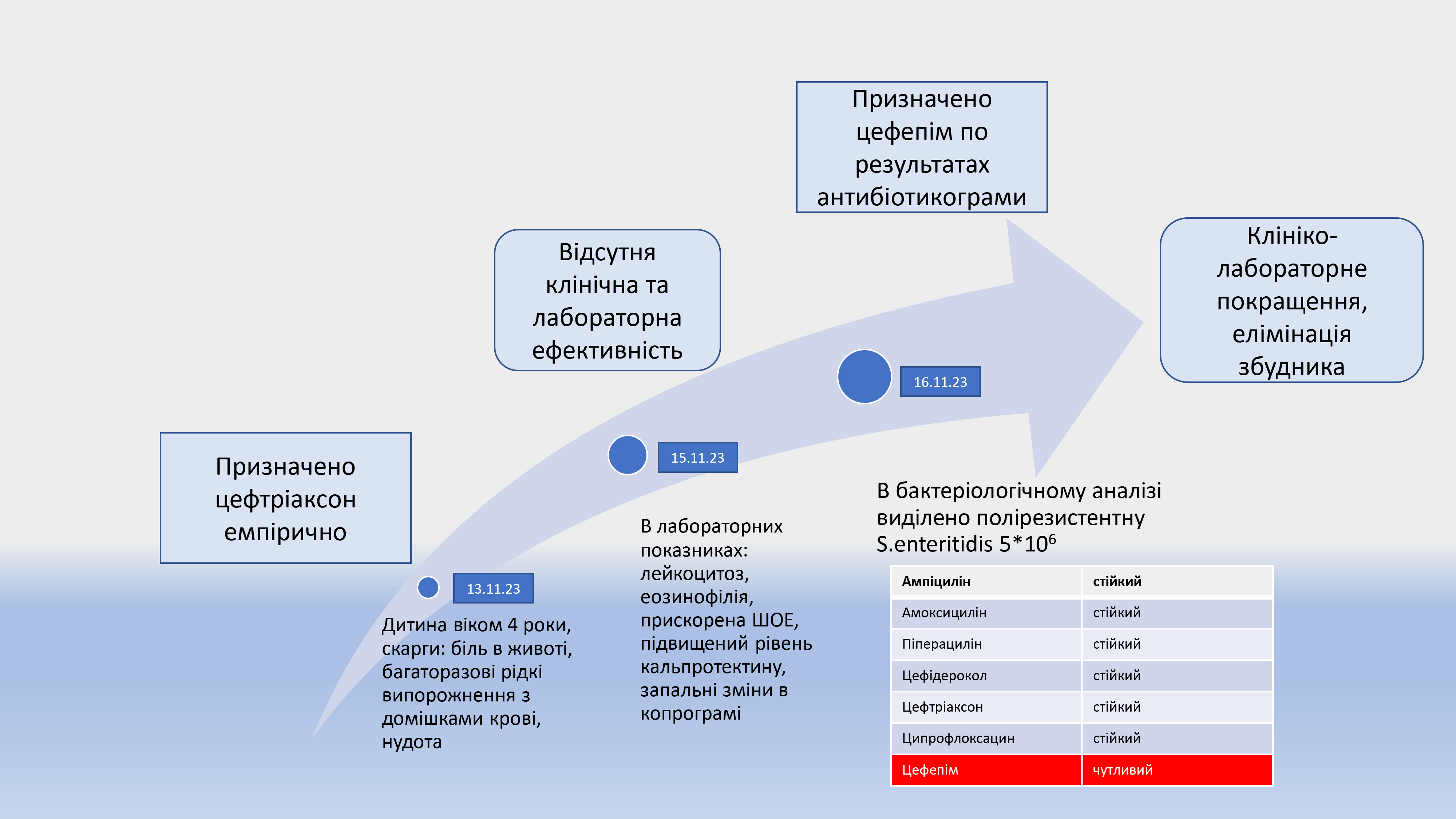
Materials and Methods. A clinical case of successful treatment of a 4-year-old child with severe intestinal involvement caused by the gastrointestinal form of salmonellosis due to a multidrug-resistant strain of Salmonella enteritidis is presented. A stepwise diagnostic approach and the rationale for selecting appropriate antibacterial therapy are described.
Case Presentation. This report describes a case of gastrointestinal salmonellosis caused by Salmonella enteritidis in a 4-year-old child. The disease had a gradual onset, presented with abdominal pain and diarrhea with mucus and blood, without fever or vomiting. Physical examination revealed moderate dehydration, periumbilical tenderness, and hepatomegaly. Laboratory findings showed leukocytosis, accelerated erythrocyte sedimentation rate (ESR), and eosinophilia. Coprological examination revealed erythrocytes, leukocytes, and neutral fat. Elevated fecal calprotectin levels and decreased fecal elastase were detected. Immunological assessment demonstrated increased IgE levels and reduced cytotoxic T lymphocytes. Endoscopic and histological examinations confirmed active eosinophilic sigmoiditis. Stool culture identified a multidrug-resistant Salmonella enteritidis strain; only fourth-generation cephalosporins were effective, resulting in complete clinical recovery.
Results and Discussion. The clinical presentation combined typical features of the gastrointestinal form of salmonellosis — bloody diarrhea, abdominal pain, hepatomegaly, and inflammatory changes in the complete blood count — with atypical manifestations, including the absence of vomiting and hyperthermia, eosinophilia, and IgE-mediated hypersensitivity. This constellation of findings suggests a mixed infectious–allergic disease course. Additionally, diagnostic markers of intestinal inflammation were identified, including a 2.6-fold increase in fecal calprotectin levels compared to age-adjusted reference values, which was corroborated by histological evidence of active intestinal inflammation in the form of eosinophilic sigmoiditis. Reduced fecal elastase levels indicated exocrine pancreatic dysfunction. An individualized therapeutic approach was applied based on antimicrobial susceptibility testing, which demonstrated multidrug resistance of the isolated Salmonella enteritidis strain. Fourth-generation cephalosporins proved to be effective in this case.
Conclusions. This case illustrates the potential development of eosinophilic sigmoiditis in children with severe salmonellosis and highlights the importance of an individualized approach to antibacterial therapy, taking antimicrobial resistance patterns into account
References
- Ryan, K. J., Ray, C. G. (2018). Sherris Medical Microbiology. McGraw Hill.
- Salmonella (non-typhoidal) (2018). World Health Organization. Available at: https://www.who.int/news-room/fact-sheets/detail/salmonella-(non-typhoidal)
- Boiko, O., Yanko, N., Pundyak, T. (2025). Epidemiological trends of salmonellosis in the cross-border regions of Ukraine and Poland (2014-2023). Bulletin of Medical and Biological Research, 69–78. https://doi.org/10.63341/bmbr/1.2025.69
- Ao, T. T., Feasey, N. A., Gordon, M. A., Keddy, K. H., Angulo, F. J., Crump, J. A. (2015). Global Burden of Invasive NontyphoidalSalmonellaDisease, 20101. Emerging Infectious Diseases, 21 (6), 941–949. https://doi.org/10.3201/eid2106.140999
- Salmonellosis – Annual Epidemiological Report for 2022 (2023). Stockholm: European Centre for Disease Prevention and Control.
- The European Union One Health 2022 Zoonoses Report (2023). EFSA Journal, 21 (12). https://doi.org/10.2903/j.efsa.2023.8442
- Guerrant, R. L., Walker, D. H., Weller, P. F. (2020). Tropical Infectious Diseases: Principles, Pathogens and Practice. Elsevier.
- Fàbrega, A., Vila, J. (2013). Salmonella enterica Serovar Typhimurium Skills To Succeed in the Host: Virulence and Regulation. Clinical Microbiology Reviews, 26 (2), 308–341. https://doi.org/10.1128/cmr.00066-12
- Santos, R. L. (2014). Pathobiology of Salmonella, Intestinal Microbiota, and the Host Innate Immune Response. Frontiers in Immunology, 5. https://doi.org/10.3389/fimmu.2014.00252
- Gullberg, R. C., Steel, J. J., Pujari, V., Rovnak, J., Crick, D. C., Perera, R. (2018). Stearoly-CoA desaturase 1 differentiates early and advanced dengue virus infections and determines virus particle infectivity. PLOS Pathogens, 14 (8), e1007261. https://doi.org/10.1371/journal.ppat.1007261
- Podavalenko, A., Malysh, N., Zadorozhna, V., Chemych, M., Biryukova, S., Chorna, I. (2021). Incidence and risk factors of salmonellosis in Ukraine. Folia Medica Cracoviensia, 61 (2), 91–102. https://doi.org/10.24425/fmc.2021.137226
- Lusta, M., Voronkova, O., Chornyi, V., Breus, A., Yesaulenko, I., Shulzhenko, D. et al. (2024). Monitoring of salmonella enterica resistance to antibiotics among children with acute intestinal infections. Eastern Ukrainian Medical Journal, 12 (4), 777–787. https://doi.org/10.21272/eumj.2024;12(4):777-787
- Su, J., Zhong, W., Liang, B., Wang, Y. (2025). Clinical characteristics and prognosis of non-typhoidal Salmonella bacteremia in children vs. adults: a retrospective study. Frontiers in Medicine, 12. https://doi.org/10.3389/fmed.2025.1597371
- Romagnani, S. (1991). Human TH1 and TH2 subsets: doubt no more. Immunology Today, 12 (8), 256–257. https://doi.org/10.1016/0167-5699(91)90120-i
- Ness, T. E., Erickson, T. A., Diaz, V., Grimes, A. B., Rochat, R., Anvari, S. et al. (2023). Pediatric Eosinophilia: A Review and Multiyear Investigation into Etiologies. The Journal of Pediatrics, 253, 232-237.e1. https://doi.org/10.1016/j.jpeds.2022.09.048
- Fitz Patrick, R. D., Noone, J. R., Cartwright, R. A., Gatti, D. M., Brosschot, T. P., Lane, J. M., Reynolds, L. A. (2024). Eosinophils respond to, but are not essential for control of an acute Salmonella enterica serovar Typhimurium infection in mice. Infection and Immunity, 92 (10). https://doi.org/10.1128/iai.00325-24
- Kramarov, S. O., Yevtushenko, V. V., Yevtushenko, О. М., Maevska, Ye. A., Babak, V. V. (2022). The problem of dehydration in pediatrics. Child`S Health, 16 (4), 296–303. https://doi.org/10.22141/2224-0551.16.4.2021.236909
- van Rheenen, P. F., Van de Vijver, E., Fidler, V. (2010). Faecal calprotectin for screening of patients with suspected inflammatory bowel disease: diagnostic meta-analysis. BMJ, 341 (1), c3369–c3369. https://doi.org/10.1136/bmj.c3369
- Jukic, A., Bakiri, L., Wagner, E. F., Tilg, H., Adolph, T. E. (2021). Calprotectin: from biomarker to biological function. Gut, 70 (10), 1978–1988. https://doi.org/10.1136/gutjnl-2021-324855
- Denysova, M., Zadorozhna, T., Bukulova, N., Archakova, T. (2021). Pathomorphological features of clinical forms of ulcerative colitis in children. Child`s health,16 (1), 1–7. https://doi.org/10.22141/2224-0551.16.1.2021.226445
- Rezultaty monitorynhu (ne)ratsionalnoho zastosuvannia antybakterialnykh preparativ za 2024 rik (2025). Tsentr hromadskoho zdorovia MOZ Ukrainy. Available at: https://www.phc.org.ua/news/rezultati-monitoringu-neracionalnogo-zastosuvannya-antibakterialnikh-preparativ-za-2024-rik
- Belousova, O. Yu. (2021). Acute Gastroenteritis in Children. Updated ESPGHAN 2020 Guidelines. Pediatrics. Eastern Europe, 1, 143–150. https://doi.org/10.34883/pi.2021.9.1.012
Downloads
Published
How to Cite
Issue
Section
License
Copyright (c) 2025 Iryna Nezgoda, Olha Naumenko, Yaroslav Demchyshyn, Olena Onofriichuk, Liudmila Starynets

This work is licensed under a Creative Commons Attribution 4.0 International License.
Our journal abides by the Creative Commons Attribution 4.0 International License copyright rights and permissions for open access journals.
Authors, who are published in this journal, agree to the following conditions:
1. The authors reserve the right to authorship of the work and pass the first publication right of this work to the journal under the terms of a Creative Commons Attribution 4.0 International License, which allows others to freely distribute the published research with the obligatory reference to the authors of the original work and the first publication of the work in this journal.
2. The authors have the right to conclude separate supplement agreements that relate to non-exclusive work distribution in the form in which it has been published by the journal (for example, to upload the work to the online storage of the journal or publish it as part of a monograph), provided that the reference to the first publication of the work in this journal is included.
3. Authors have the right to store the final accepted version of the article in an institutional, thematic, or any other repository to ensure visibility and accessibility.
