The value of computer reconstruction in the treatment of intra-articular fractures of the distal part of the humerus
DOI:
https://doi.org/10.15587/2519-4798.2024.308333Keywords:
computed tomography, preoperative planning, distal humerus fracture, 3D printing, 3D modelling, fracture, treatmentAbstract
The aim of the work was to study the role of computer reconstruction and additive technologies in the planning of surgical treatment of intra-articular fractures of the distal part of the humerus in the early post-traumatic period.
Materials and methods: the results of treatment of 44 patients with fractures of the distal part of the humerus aged from 22 to 65 years were studied.
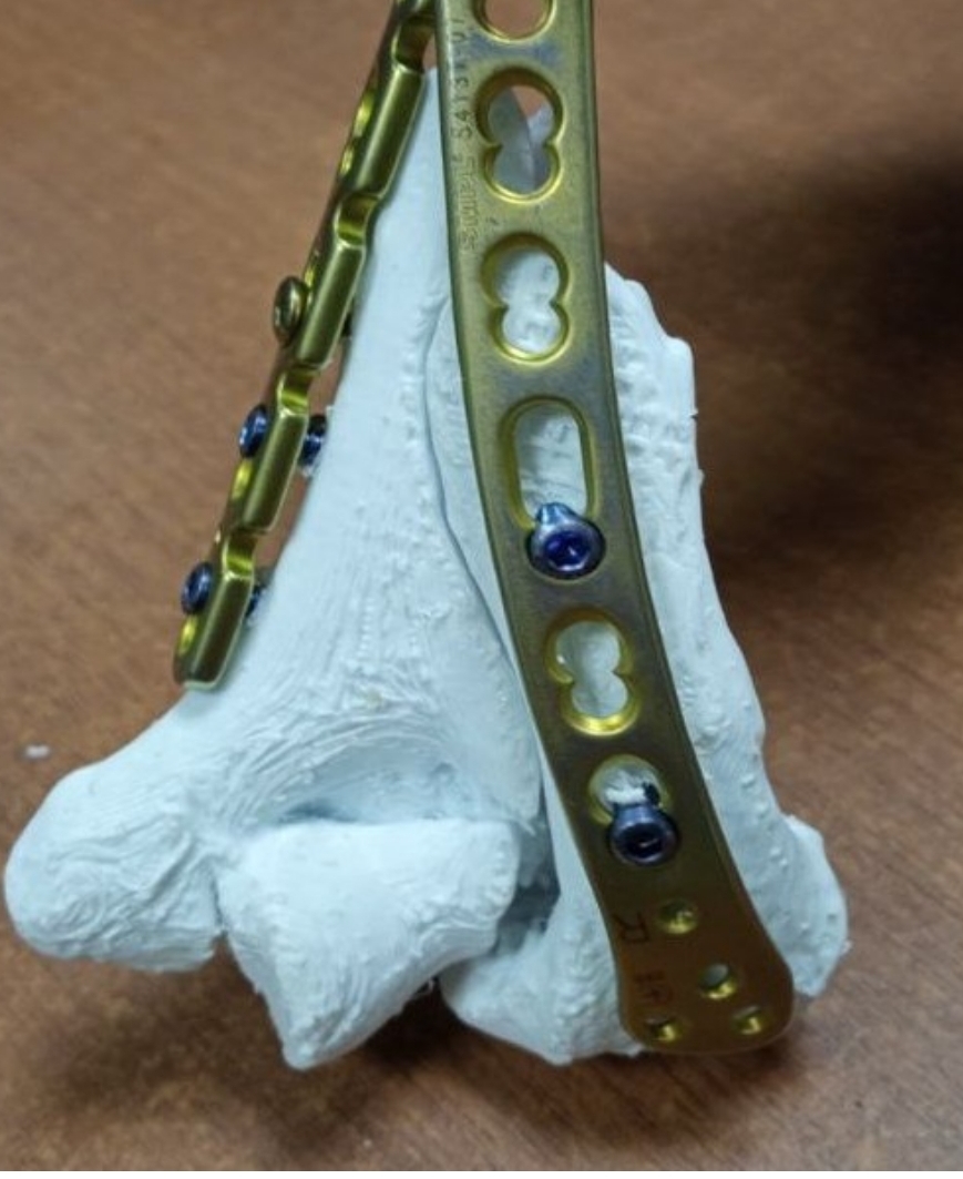
All patients underwent MSCT in the preoperative period. In 45.5 % (20) cases, in order to increase the effectiveness of preoperative planning, simulation osteosynthesis was performed on an individual printed model of the damaged bone.
After completion of preoperative planning, all patients underwent osseous metallo-osteosynthesis in the first week after receiving the injury. In order to assess the effectiveness of the application of visualization techniques, a survey of the operating team was carried out regarding the informativeness of the conducted instrumental studies.
Results. The average duration of surgical intervention among patients in the clinical group with standard preoperative planning was 105.9 ± 9.15 minutes. The average results of the survey of the surgical team after the surgical interventions in the specified clinical group were 21.2 ± 2.8 points.
The average duration of surgery among patients of the second clinical group, whose preoperative planning included not only the assessment of MSCT results of the damaged segment, but also simulated osteosynthesis with the selection and application of optimal metal fixators, was 54.6 ± 7.14 minutes. The average results of the survey of the surgical team were 31.2 ± 1.7 points.
Conclusions. The use of a combination of visualization technologies and 3D printing allows to reduce the time the patient spends in the operating room and increase the effectiveness of preoperative planning.
Performing simulated osteosynthesis in the preoperative period allows you to develop an individual technique of repositioning bone fragments and use the optimal standard size of the cortical metal fixator with the distribution of the most favourable compression points of bone fragments
References
- Folco, G., Messina, C., Gitto, S., Fusco, S., Serpi, F., Zagarella, A. et al. (2024). CT Arthrography of the Elbow: What Radiologists Should Know. Tomography, 10 (3), 415–427. https://doi.org/10.3390/tomography10030032
- Lubet, A., Renaux-Petel, M., Delbreilh, L., Liard-Zmuda, A., Auble, A., Payen, M. (2024). Conception and validation of A 3d printed learning model of supra condylar fracture of children. Heliyon, 10 (10), e30741. https://doi.org/10.1016/j.heliyon.2024.e30741
- Lal, H., Patralekh, M. K. (2018). 3D printing and its applications in orthopaedic trauma: A technological marvel. Journal of Clinical Orthopaedics and Trauma, 9 (3), 260–268. https://doi.org/10.1016/j.jcot.2018.07.022
- Moursy, M., Wegmann, K., Wichlas, F., Tauber, M. (2020). Distal humerus fracture in patients over 70 years of age: results of open reduction and internal fixation. Archives of Orthopaedic and Trauma Surgery, 142 (1), 157–164. https://doi.org/10.1007/s00402-020-03664-4
- Lawan Abdou, A., El aissaoui, T., Lachkar, A., Abdeljaouad, N., Yacoubi, H. (2024). A Partial Frontal Fracture of the Humeral Trochlea: A Case Report. Cureus. https://doi.org/10.7759/cureus.56640
- Vauclair, F., Goetti, P., Nguyen, N. T. V., Sanchez-Sotelo, J. (2020). Distal humerus nonunion: evaluation and management. EFORT Open Reviews, 5 (5), 289–298. https://doi.org/10.1302/2058-5241.5.190050
- Grunert, R., Winkler, D., Frank, F., Moebius, R., Kropla, F., Meixensberger, J. et al. (2023). 3D-printing of the elbow in complex posttraumatic elbow-stiffness for preoperative planning, surgery-simulation and postoperative control. 3D Printing in Medicine, 9 (1). https://doi.org/10.1186/s41205-023-00191-x
- Przyklenk, A., Hackl, M., Iuga, A.-I., Leschinger, T., Maintz, D., Harbrecht, A. et al. (2023). Computed tomography-based angle measurements of the sagittal capitulum and trochlea position in relation to the humeral shaft. Surgical and Radiologic Anatomy, 45 (5), 571–580. https://doi.org/10.1007/s00276-023-03118-7
- Sonnow, L., Salimova, N., Behrendt, L., Wacker, F. K., Örgel, M., Plagge, J., Weidemann, F. (2023). Photon-counting CT of elbow joint fractures: image quality in a simulated post-trauma setting with off-center positioning. European Radiology Experimental, 7 (1). https://doi.org/10.1186/s41747-023-00329-w
- Emery, K. H., Zingula, S. N., Anton, C. G., Salisbury, S. R., Tamai, J. (2015). Pediatric elbow fractures: a new angle on an old topic. Pediatric Radiology, 46 (1), 61–66. https://doi.org/10.1007/s00247-015-3439-0
- Kraus, R., Dresing, K. (2023). Rational Usage of Fracture Imaging in Children and Adolescents. Diagnostics, 13 (3), 538. https://doi.org/10.3390/diagnostics13030538
- Wong, W.-S.-Y., Sim, C., Tay, Z. Q., Yeap, P. M., Seah, R. B. (2024). Targeted four-dimensional computerized tomography scans for elbow disorders: a literature review and refinement of existing technique with two exemplar cases. JSES International, 8 (2), 378–383. https://doi.org/10.1016/j.jseint.2023.11.014
- Mogharrabi, B., Cabrera, A., Chhabra, A. (2022). 3D isotropic spine echo MR imaging of elbow: How it helps surgical decisions. European Journal of Radiology Open, 9, 100410. https://doi.org/10.1016/j.ejro.2022.100410
- Kononenko, S. V., Pelypenko, О. V. (2022). Anatomical features of the soft tissue structures of the proximal humeral diaphysis revealed by the magnetic resonance imaging. Bulletin of Problems Biology and Medicine, 1 (2), 184. https://doi.org/10.29254/2077-4214-2022-2-1-164-184-189
- Calvo-Haro, J. A., Pascau, J., Mediavilla-Santos, L., Sanz-Ruiz, P., Sánchez-Pérez, C., Vaquero-Martín, J., Perez-Mañanes, R. (2021). Conceptual evolution of 3D printing in orthopedic surgery and traumatology: from “do it yourself” to “point of care manufacturing.” BMC Musculoskeletal Disorders, 22 (1). https://doi.org/10.1186/s12891-021-04224-6
- Andrés-Cano, P., Calvo-Haro, J. A., Fillat-Gomà, F., Andrés-Cano, I., Perez-Mañanes, R. (2021). Role of the orthopaedic surgeon in 3D printing: current applications and legal issues for a personalized medicine. Revista Española de Cirugía Ortopédica y Traumatología (English Edition), 65 (2), 138–151. https://doi.org/10.1016/j.recote.2021.01.001
- Teo, A. Q. A., Ng, D. Q. K., Lee, P., O’neill, G. K. (2021). Point-of-care 3D printing: A feasibility study of using 3D printing for orthopaedic trauma. Injury, 52 (11), 3286–3292. https://doi.org/10.1016/j.injury.2021.02.041
- Mendonça, C. J. A., Guimarães, R. M. da R., Pontim, C. E., Gasoto, S. C., Setti, J. A. P., Soni, J. F., Schneider, B. (2023). An Overview of 3D Anatomical Model Printing in Orthopedic Trauma Surgery. Journal of Multidisciplinary Healthcare, 16, 875–887. https://doi.org/10.2147/jmdh.s386406
- Kulenova, N. A., Dogadkin, S., Azamatov, N., Sadenova, A., Beisekenov, N. A., Shaimardanov, Z. K. et al. (2022). Modeling and manufacturing of individual implants for traumatology and orthopedics. Chemical Engineering Transactions. https://doi.org/10.3303/CET2294132
- Suffo, M., López-Marín, C., Revenga, C., Andrés-Cano, P. (2024). Polyetherimide in 3D printing: Pioneering non-metallic solutions for personalized orthopedic and traumatology hip prosthetics. Results in Engineering, 23, 102372. https://doi.org/10.1016/j.rineng.2024.102372
Downloads
Published
How to Cite
Issue
Section
License
Copyright (c) 2024 Olexandr Kovalov, Olexandr Pelypenko, Serhii Kononenko, Svitlana Pavlenko, Serghii Malyk

This work is licensed under a Creative Commons Attribution 4.0 International License.
Our journal abides by the Creative Commons CC BY copyright rights and permissions for open access journals.
Authors, who are published in this journal, agree to the following conditions:
1. The authors reserve the right to authorship of the work and pass the first publication right of this work to the journal under the terms of a Creative Commons CC BY, which allows others to freely distribute the published research with the obligatory reference to the authors of the original work and the first publication of the work in this journal.
2. The authors have the right to conclude separate supplement agreements that relate to non-exclusive work distribution in the form in which it has been published by the journal (for example, to upload the work to the online storage of the journal or publish it as part of a monograph), provided that the reference to the first publication of the work in this journal is included.