Порівняльна оцінка загальновживаних таблиць пошуку кольорів для визначення ключових показників ефективності візуалізації даних перфузійних карт
DOI:
https://doi.org/10.15587/2706-5448.2026.352787Ключові слова:
схема відображення кольорів, сприйняття кольору, візуалізація кольору, гемодинамічні параметри, перфузійно-зважені зображенняАнотація
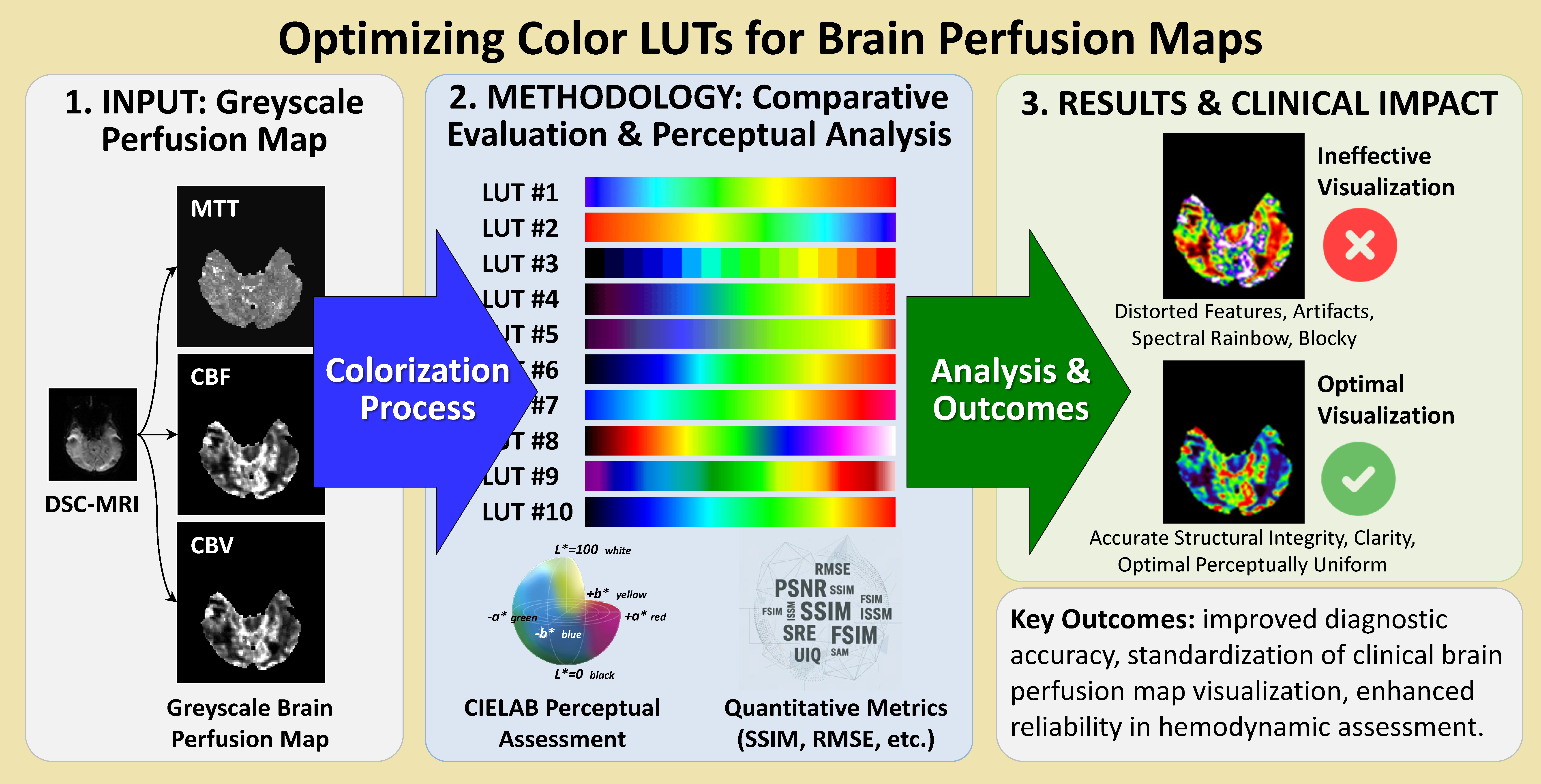
Об’єктом даного дослідження є таблиці пошуку кольорів, які найчастіше використовують під час візуалізації перфузійних карт для оцінки гемодинамічних параметрів мозку. Проблема полягає в тому, що такі колірні схеми значно відрізняються за кількістю кольорів, їх розподілом і правилами переведення даних сірошкальних зображень у кольорові. У наслідок одна й та сама перфузійна карта може виглядати по-різному, залежно від використаної схеми, що ускладнює візуальну оцінку гемодинамічних параметрів і значно впливає на точність їх інтерпретації.
Дане дослідження містить комплексний аналіз десяти загально використовуваних таблиць пошуку кольорів, які поширені під час візуалізації перфузійних карт. Надаються результати оцінювання як на основі безпосереднього аналізу даних самих схем, так і за даними перфузійних карт реальних клінічних випадків. Серед кількісних показників – RMSE, PSNR, SSIM, FSIM, ISSM, SRE, SAM та UIQ. Колірний простір CIELAB використовують для оцінювання сприйняття змін у кольорі між суміжними елементами таблиць пошуку кольорів. Він також використовується для аналізу взаємозв’язку між локальними змінами інтенсивності та можливостями сприйняття змін у кольорі в перфузіних картах до та після їх колоризації. Аналіз показує, що вибір схем таблиць пошуку кольорів має вирішальне значення для збереження змін інтенсивності показників і структурної цілісності. Спектральні райдужні та блочні за структурою схеми поступаються іншим щодо ефективності через спотворення структурних особливостей.
Результати можуть бути застосовані на практиці під час візуалізації даних перфузійних карт у медичному програмному забезпеченні для оцінки ключових гемодинамічних параметрів, таких як об’єм крові, кровотік і середній час проходження. Крім того, результати можуть бути корисними для стандартизації та вибору оптимальних схем таблиць пошуку кольорів у клінічній практиці, а також для перевірки алгоритмів, які використовуються для розрахунку перфузійних карт під час розробки медичного програмного забезпечення.
Посилання
- Putowski, Z., Bakker, J., Kattan, E., Hernández, G., Ait-Oufella, H., Szczeklik, W., Guerci, P. (2025). Tissue perfusion as the ultimate target of hemodynamic interventions in the perioperative period. Journal of Clinical Anesthesia, 107, 112009. https://doi.org/10.1016/j.jclinane.2025.112009
- Lipiński, S. (2024). Creation of a Simulated Sequence of Dynamic Susceptibility Contrast–Magnetic Resonance Imaging Brain Scans as a Tool to Verify the Quality of Methods for Diagnosing Diseases Affecting Brain Tissue Perfusion. Computation, 12 (3), 54. https://doi.org/10.3390/computation12030054
- Sobhan, R., Gkogkou, P., Johnson, G., Cameron, D. (2023). Model-based deconvolution for DSC-MRI: A comparison of accuracy, precision, and computational complexity of parametric transit time distributions. https://doi.org/10.1101/2023.02.12.528216
- Sorokina, V. V., Alkhimova, S. M. (2024). Analiz zastosuvannia tablyts vidpovidnosti koloriv dlia vizualizatsii danykh perfuziinykh kart. World Ways and Methods of Improving Outdated Theories and Trends, 380–385. Available at: https://isg-konf.com/world-ways-and-methods-of-improving-outdated-theories-and-trends/
- Căiniceanu, A.-M.-A., Alexa, F. (2025). Application of Color Theory in Graphic Design. 2025 18th International Conference on Engineering of Modern Electric Systems (EMES). IEEE 1–6. https://doi.org/10.1109/emes65692.2025.11045619
- Furmanová, K., Kozlíková, B., Höllt, T., Gröller, M. E., Preim, B., Raidou, R. G. (2025). BioMedical Visualization. Synthesis Lectures on Visualization. Springer Nature Switzerland. https://doi.org/10.1007/978-3-031-66789-3
- Preim, B., Oeltze, S., Mlejnek, M., Groeller, E., Hennemuth, A., Behrens, S. (2009). Survey of the Visual Exploration and Analysis of Perfusion Data. IEEE Transactions on Visualization and Computer Graphics, 15 (2), 205–220. https://doi.org/10.1109/tvcg.2008.95
- Garrison, L. A., Kolesar, I., Viola, I., Hauser, H., Bruckner, S. (2022). Trends & Opportunities in Visualization for Physiology: A Multiscale Overview. Computer Graphics Forum, 41 (3), 609–643. https://doi.org/10.1111/cgf.14575
- Crameri, F., Shephard, G. E., Heron, P. J. (2020). The misuse of colour in science communication. Nature Communications, 11 (1). https://doi.org/10.1038/s41467-020-19160-7
- Moreland, K. (2016). Why We Use Bad Color Maps and What You Can Do About It. Electronic Imaging, 28 (16), 1–6. https://doi.org/10.2352/issn.2470-1173.2016.16.hvei-133
- Reda, K. (2023). Rainbow Colormaps: What are They Good and Bad for? IEEE Transactions on Visualization and Computer Graphics, 29 (12), 5496–5510. https://doi.org/10.1109/tvcg.2022.3214771
- Liu, Y., Heer, J. (2018). Somewhere Over the Rainbow. Proceedings of the 2018 CHI Conference on Human Factors in Computing Systems, 1–12. https://doi.org/10.1145/3173574.3174172
- Stoelzle, M., Stein, L. (2021). Rainbow color map distorts and misleads research in hydrology – guidance for better visualizations and science communication. Hydrology and Earth System Sciences, 25 (8), 4549–4565. https://doi.org/10.5194/hess-25-4549-2021
- Silva, S., Sousa Santos, B., Madeira, J. (2011). Using color in visualization: A survey. Computers & Graphics, 35 (2), 320–333. https://doi.org/10.1016/j.cag.2010.11.015
- Sibrel, S. C., Rathore, R., Lessard, L., Schloss, K. B. (2020). The relation between color and spatial structure for interpreting colormap data visualizations. Journal of Vision, 20 (12), 7. https://doi.org/10.1167/jov.20.12.7
- Zabala-Travers, S., Choi, M., Cheng, W.-C., Badano, A. (2015). Effect of color visualization and display hardware on the visual assessment of pseudocolor medical images. Medical Physics, 42 (6), 2942–2954. https://doi.org/10.1118/1.4921125
- Nuñez, J. R., Anderton, C. R., Renslow, R. S. (2018). Optimizing colormaps with consideration for color vision deficiency to enable accurate interpretation of scientific data. PLOS ONE, 13 (7), e0199239. https://doi.org/10.1371/journal.pone.0199239
- Ware, C., Turton, T. L., Bujack, R., Samsel, F., Shrivastava, P., Rogers, D. H. (2019). Measuring and Modeling the Feature Detection Threshold Functions of Colormaps. IEEE Transactions on Visualization and Computer Graphics, 25 (9), 2777–2790. https://doi.org/10.1109/tvcg.2018.2855742
- Sollmann, N., Fuderer, M., Crameri, F., Weingärtner, S., Baeßler, B., Gulani, V. et al. (2024). Color Maps: Facilitating the Clinical Impact of Quantitative MRI. Journal of Magnetic Resonance Imaging, 61 (4), 1572–1579. https://doi.org/10.1002/jmri.29573
- deSouza, N. M., Fuderer, M., Sollmann, N., Wichtmann, B. D., Golay, X. (2025). Colour map displays. Insights into Imaging, 16 (1). https://doi.org/10.1186/s13244-025-01970-2
- Qutbi, M. (2024). Quantitative Performance Evaluation of Commonly Used Colormaps for Image Display in Myocardial Perfusion Imaging: Analysis based on Perceptual Metrics. Molecular Imaging and Radionuclide Therapy, 33 (2), 94–105. https://doi.org/10.4274/mirt.galenos.2024.34711
- The Cancer Genome Atlas Program (TCGA). Cancer.gov. Available at: http://cancergenome.nih.gov/
- Jain, R., Poisson, L., Narang, J., Gutman, D., Scarpace, L., Hwang, S. N. et al. (2013). Genomic Mapping and Survival Prediction in Glioblastoma: Molecular Subclassification Strengthened by Hemodynamic Imaging Biomarkers. Radiology, 267 (1), 212–220. https://doi.org/10.1148/radiol.12120846
- Alkhimova, S., Krenevych, A. (2019). Brain tissues segmentation on mr perfusion images using CUSUM filter for boundary pixels. International Journal of Computing, 18 (2), 127–134. https://doi.org/10.47839/ijc.18.2.1411
- Alkhimova, S. (2019). CUSUM Filter for Brain Segmentation on DSC Perfusion MR Head Scans with Abnormal Brain Anatomy. Proceedings of the 2019 International Conference on Intelligent Medicine and Image Processing, 43–47. https://doi.org/10.1145/3332340.3332357
- Kudo, K., Sasaki, M., Yamada, K., Terae, S., Tha, K. K., Yoshida, Y., Miyasaka, K. (2006). Evaluation and minimization of the difference in MR perfusion maps among software. Proceedings of the 14th Annual Meeting of ISMRM, 3558. Available at: https://cds.ismrm.org/protected/06MProceedings/PDFfiles/03558.pdf
- Kudo, K. (2005). Acute Stroke Imaging Standardization Group Japan recommended standard LUT (a-LUT) for perfusion color maps.
- Schanda, J. (Ed.) (2007). Colorimetry: understanding the CIE system. John Wiley & Sons. https://doi.org/10.1002/9780470175637
- Sara, U., Akter, M., Uddin, M. S. (2019). Image Quality Assessment through FSIM, SSIM, MSE and PSNR – A Comparative Study. Journal of Computer and Communications, 7 (3), 8–18. https://doi.org/10.4236/jcc.2019.73002
- Sampat, M. P., Zhou Wang, Gupta, S., Bovik, A. C., Markey, M. K. (2009). Complex Wavelet Structural Similarity: A New Image Similarity Index. IEEE Transactions on Image Processing, 18 (11), 2385–2401. https://doi.org/10.1109/tip.2009.2025923
- Lin Zhang, Lei Zhang, Xuanqin Mou, Zhang, D. (2011). FSIM: A Feature Similarity Index for Image Quality Assessment. IEEE Transactions on Image Processing, 20 (8), 2378–2386. https://doi.org/10.1109/tip.2011.2109730
- Aljanabi, M. A., Hussain, Z. M., Shnain, N. A. A., Lu, S. F. (2019). Design of a hybrid measure for image similarity: a statistical, algebraic, and information-theoretic approach. European Journal of Remote Sensing, 52, 2–15. https://doi.org/10.1080/22797254.2019.1628617
- Nordio, A., Chiasserini, C.-F., Viterbo, E. (2009). Signal Reconstruction Errors in Jittered Sampling. IEEE Transactions on Signal Processing, 57 (12), 4711–4718. https://doi.org/10.1109/tsp.2009.2027404
- Yuhas, R. H., Goetz, A. F., Boardman, J. W. (1992). Discrimination among semi-arid landscape endmembers using the spectral angle mapper (SAM) algorithm. JPL, Summaries of the Third Annual JPL Airborne Geoscience Workshop. Volume 1: AVIRIS Workshop. Available at: https://ntrs.nasa.gov/citations/19940012193
- Zhou Wang, Bovik, A. C. (2002). A universal image quality index. IEEE Signal Processing Letters, 9 (3), 81–84. https://doi.org/10.1109/97.995823
##submission.downloads##
Опубліковано
Як цитувати
Номер
Розділ
Ліцензія
Авторське право (c) 2026 Svitlana Alkhimova, Viktoriia Sorokina, Illia Kabala

Ця робота ліцензується відповідно до Creative Commons Attribution 4.0 International License.
Закріплення та умови передачі авторських прав (ідентифікація авторства) здійснюється у Ліцензійному договорі. Зокрема, автори залишають за собою право на авторство свого рукопису та передають журналу право першої публікації цієї роботи на умовах ліцензії Creative Commons CC BY. При цьому вони мають право укладати самостійно додаткові угоди, що стосуються неексклюзивного поширення роботи у тому вигляді, в якому вона була опублікована цим журналом, але за умови збереження посилання на першу публікацію статті в цьому журналі.