Оцінка антимікробної активності біоматеріалів на основі альгінату та декаметоксину щодо Staphylococcus aureus та Escherichia coli
DOI:
https://doi.org/10.15587/2519-8025.2023.298594Ключові слова:
антимікробні біоматеріали, S.aureus, E.coli, антисептики, декаметоксин, альгінат кальцію, антибіотикорезистентністьАнотація
Мета – вивчення антимікробної активності нових біоматеріалів на основі декаметоксину та комерційно доступних ранових покриттів щодо референтних та клінічних штамів S.aureus та E.coli.
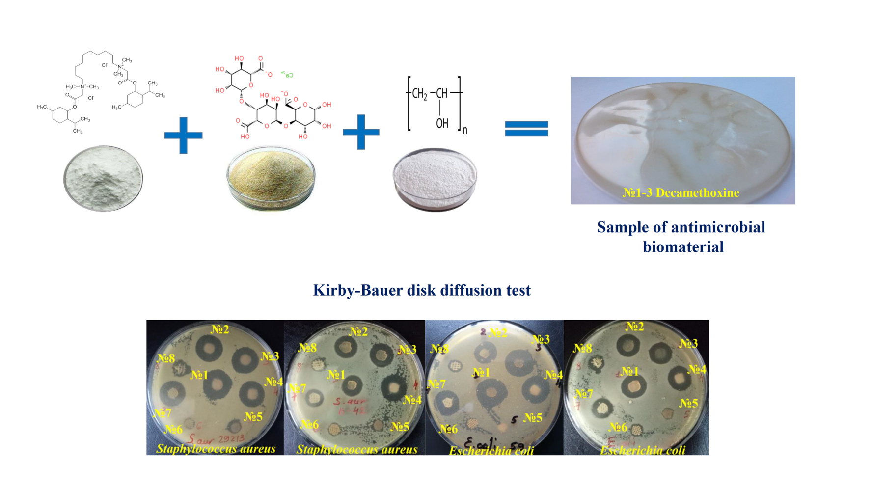
Матеріали і методи. Для дослідження використовували розроблені біоматеріали з декаметоксином 0.05 % (ДКМ № 1-3 ) та ранові пов’язки з вмістом антисептиків Suprasorb®, SILVERCEL®, Urgotul SSD®, GUANPOLISEPT®, Bétadine®. Протимікробні властивості вивчали методом дискової дифузії з реєстрацією та порівнянням діаметрів зон затримки росту (ЗЗР).
Результати. По відношенню до S.aureus АТСС 25923 встановлено значно вищу антимікробну активність біоматеріалів з ДКМ у порівнянні з срібло- та йод-вмісними антимікробними рановими покриттями у 1.97-2.11 (p <0.001) та 1.73-1.86 рази (p <0.001) відповідно. Подібною активністю щодо S.aureus АТСС 25923 володіли усі три зразки з ДКМ (ЗЗР - від 21.98±0.18 до 23.58±0.26 мм) та ранові покриття Suprasorb® (19.31±0.17 мм), Guanpolisept® (19.13±0.12 мм). Таку тенденцію також виявили щодо клінічних штамів стафілококів. Високий рівень активності щодо E.coli ATCC 25922 показали біоматеріали № 1-3 ДКМ (ЗЗР - від 19.01±0.33 до 21.54±0.23 мм), Guanpolisept® (18.74±0.12 мм) та Suprasorb® (18.43±0.13 мм). Клінічні штами E.coli проявляли більшу толерантність щодо антимікробних біоматеріалів: різниця середніх значень між ЗЗР референтного та ЗЗР клінічних штамів E.coli була достовірною для всіх біоматеріалів (p <0.001) Найбільш ефективними були біоматеріали ДКМ № 1-3 (ЗЗР - від 15.58±0.25 до 16.41±0.16 мм), а також полігексанід-вмісна пов’язка Suprasorb® (15.82±0.31 мм).
Висновки. Найвищою антистафілококовою активністю володіють біоматеріали на основі декаметоксину № 1, № 2, № 3, Suprasorb®, Guanpolisept® та Bétadine®. Найсильнішу дію на референтні та клінічні штами E.coli проявляють біоматеріали з декаметоксином № 1-3, Suprasorb® та Guanpolisept®
Посилання
- Guiomar, A. J., Urbano, A. M. (2022). Polyhexanide-Releasing Membranes for Antimicrobial Wound Dressings: A Critical Review. Membranes, 12 (12), 1281. doi: https://doi.org/10.3390/membranes12121281
- Liang, Y., Liang, Y., Zhang, H., Guo, B. (2022). Antibacterial biomaterials for skin wound dressing. Asian Journal of Pharmaceutical Sciences, 17 (3), 353–384. doi: https://doi.org/10.1016/j.ajps.2022.01.001
- Boateng, J., Catanzano, O. (2015). Advanced Therapeutic Dressings for Effective Wound Healing – A Review. Journal of Pharmaceutical Sciences, 104 (11), 3653–3680. doi: https://doi.org/10.1002/jps.24610
- Norouzi, M., Boroujeni, S. M., Omidvarkordshouli, N., Soleimani, M. (2015). Advances in Skin Regeneration: Application of Electrospun Scaffolds. Advanced Healthcare Materials, 4 (8), 1114–1133. doi: https://doi.org/10.1002/adhm.201500001
- Pahlevanzadeh, F., Setayeshmehr, M., Bakhsheshi-Rad, H. R., Emadi, R., Kharaziha, M., Poursamar, S. A., Ismail, A. F., Sharif, S., Chen, X., Berto, F. (2022). A Review on Antibacterial Biomaterials in Biomedical Applications: From Materials Perspective to Bioinks Design. Polymers, 14 (11), 2238. doi: https://doi.org/10.3390/polym14112238
- Sam, S., Joseph, B., Thomas, S. (2023). Exploring the antimicrobial features of biomaterials for biomedical applications. Results in Engineering, 17, 100979. doi: https://doi.org/10.1016/j.rineng.2023.100979
- Yu, R., Zhang, H., Guo, B. (2021). Conductive Biomaterials as Bioactive Wound Dressing for Wound Healing and Skin Tissue Engineering. Nano-Micro Letters, 14 (1). doi: https://doi.org/10.1007/s40820-021-00751-y
- Dodero, A., Scarfi, S., Pozzolini, M., Vicini, S., Alloisio, M., Castellano, M. (2019). Alginate-Based Electrospun Membranes Containing ZnO Nanoparticles as Potential Wound Healing Patches: Biological, Mechanical, and Physicochemical Characterization. ACS Applied Materials & Interfaces, 12 (3), 3371–3381. doi: https://doi.org/10.1021/acsami.9b17597
- Da Silva, J., Leal, E. C., Carvalho, E., Silva, E. A. (2023). Innovative Functional Biomaterials as Therapeutic Wound Dressings for Chronic Diabetic Foot Ulcers. International Journal of Molecular Sciences, 24 (12), 9900. doi: https://doi.org/10.3390/ijms24129900
- Simões, D., Miguel, S. P., Ribeiro, M. P., Coutinho, P., Mendonça, A. G., Correia, I. J. (2018). Recent advances on antimicrobial wound dressing: A review. European Journal of Pharmaceutics and Biopharmaceutics, 127, 130–141. doi: https://doi.org/10.1016/j.ejpb.2018.02.022
- Falk, N. A. (2019). Surfactants as Antimicrobials: A Brief Overview of Microbial Interfacial Chemistry and Surfactant Antimicrobial Activity. Journal of Surfactants and Detergents, 22(5), 1119–1127. doi: https://doi.org/10.1002/jsde.12293
- Babalska, Z. Ł., Korbecka-Paczkowska, M., Karpiński, T. M. (2021). Wound Antiseptics and European Guidelines for Antiseptic Application in Wound Treatment. Pharmaceuticals, 14 (12), 1253. doi: https://doi.org/10.3390/ph14121253
- Joyce, K., Fabra, G. T., Bozkurt, Y., Pandit, A. (2021). Bioactive potential of natural biomaterials: identification, retention and assessment of biological properties. Signal Transduction and Targeted Therapy, 6 (1). doi: https://doi.org/10.1038/s41392-021-00512-8
- EUCAST disk diffusion test methodology (2015). European committee on antimicrobial susceptibility testing (EUCAST). Available at: https://www.eucast.org/ast_of_bacteria/disk_diffusion_methodology/ Last accessed: 12.08.2015
- Routine and extended internal quality control for MIC determination and disk diffusion as recommended by EUCAST version 12.0 (2022). European Committee on Antimicrobial Susceptibility Testing; Växjö.
- Matuschek, E., Longshaw, C., Takemura, M., Yamano, Y., Kahlmeter, G. (2022). Cefiderocol: EUCAST criteria for disc diffusion and broth microdilution for antimicrobial susceptibility testing. Journal of Antimicrobial Chemotherapy, 77 (6), 1662–1669. doi: https://doi.org/10.1093/jac/dkac080
- Antimicrobial Susceptibility Testing, EUCAST Disk Diffusion Method, Version 11.0 (2023). The European Committee on Antimicrobial Susceptibility Testing (EUCAST). Available at: https://www.eucast.org/fileadmin/src/media/PDFs/EUCAST_files/Disk_test_documents/2023_manuals/Manual_v_11.0_EUCAST_Disk_Test_2023.pdf Last accessed: 10.01.2023
- Chambers, H. F., DeLeo, F. R. (2009). Waves of resistance: Staphylococcus aureus in the antibiotic era. Nature Reviews Microbiology, 7 (9), 629–641. doi: https://doi.org/10.1038/nrmicro2200
- Ejaz, M., Syed, M. A., Jackson, C. R., Sharif, M., Faryal, R. (2023). Epidemiology of Staphylococcus aureus Non-Susceptible to Vancomycin in South Asia. Antibiotics, 12 (6), 972. doi: https://doi.org/10.3390/antibiotics12060972
- Reich, P. J., Boyle, M. G., Hogan, P. G., Johnson, A. J., Wallace, M. A., Elward, A. M. et al. (2016). Emergence of community-associated methicillin-resistant Staphylococcus aureus strains in the neonatal intensive care unit: an infection prevention and patient safety challenge. Clinical Microbiology and Infection, 22 (7), 645.e1–645.e8. doi: https://doi.org/10.1016/j.cmi.2016.04.013
- Yang, E. S., Tan, J., Eells, S., Rieg, G., Tagudar, G., Miller, L. G. (2010). Body site colonization in patients with community-associated methicillin-resistant Staphylococcus aureus and other types of S. aureus skin infections. Clinical Microbiology and Infection, 16 (5), 425–431. doi: https://doi.org/10.1111/j.1469-0691.2009.02836.x
- Linz, M. S., Mattappallil, A., Finkel, D., Parker, D. (2023). Clinical Impact of Staphylococcus aureus Skin and Soft Tissue Infections. Antibiotics, 12 (3), 557. doi: https://doi.org/10.3390/antibiotics12030557
- Esposito, S., Blasi, F., Curtis, N., Kaplan, S., Lazzarotto, T., Meschiari, M. et al. (2023). New Antibiotics for Staphylococcus aureus Infection: An Update from the World Association of Infectious Diseases and Immunological Disorders (WAidid) and the Italian Society of Anti-Infective Therapy (SITA). Antibiotics, 12 (4), 742. doi: https://doi.org/10.3390/antibiotics12040742
- Upreti, N., Rayamajhee, B., Sherchan, S. P., Choudhari, M. K., Banjara, M. R. (2018). Prevalence of methicillin resistant Staphylococcus aureus, multidrug resistant and extended spectrum β-lactamase producing gram negative bacilli causing wound infections at a tertiary care hospital of Nepal. Antimicrobial Resistance & Infection Control, 7 (1). doi: https://doi.org/10.1186/s13756-018-0408-z
- Tefera, S., Awoke, T., Mekonnen, D. (2021). Methicillin and Vancomycin Resistant Staphylococcus aureus and Associated Factors from Surgical Ward Inpatients at Debre Markos Referral Hospital, Northwest Ethiopia. Infection and Drug Resistance, 14, 3053–3062. doi: https://doi.org/10.2147/idr.s324042
- Braz, V. S., Melchior, K., Moreira, C. G. (2020). Escherichia coli as a Multifaceted Pathogenic and Versatile Bacterium. Frontiers in Cellular and Infection Microbiology, 10. doi: https://doi.org/10.3389/fcimb.2020.548492
- Wilcox, M. H., Dryden, M. (2021). Update on the epidemiology of healthcare-acquired bacterial infections: focus on complicated skin and skin structure infections. Journal of Antimicrobial Chemotherapy, 76 (4), iv2–iv8. doi: https://doi.org/10.1093/jac/dkab350
- Puca, V., Marulli, R. Z., Grande, R., Vitale, I., Niro, A., Molinaro, G. et al. (2021). Microbial Species Isolated from Infected Wounds and Antimicrobial Resistance Analysis: Data Emerging from a Three-Years Retrospective Study. Antibiotics, 10 (10), 1162. doi: https://doi.org/10.3390/antibiotics10101162
- Urase, T., Okazaki, M., Tsutsui, H. (2020). Prevalence of ESBL-producing Escherichia coli and carbapenem-resistant Enterobacteriaceae in treated wastewater: a comparison with nosocomial infection surveillance. Journal of Water and Health, 18 (6), 899–910. doi: https://doi.org/10.2166/wh.2020.014
- Tian, X., Sun, S., Jia, X., Zou, H., Li, S., Zhang, L. (2018). Epidemiology of and risk factors for infection with extended-spectrum β-lactamase-producing carbapenem-resistant Enterobacteriaceae: results of a double case–control study. Infection and Drug Resistance, 11, 1339–1346. doi: https://doi.org/10.2147/idr.s173456
- Kramer, A., Dissemond, J., Kim, S., Willy, C., Mayer, D., Papke, R. et al. (2017). Consensus on Wound Antisepsis: Update 2018. Skin Pharmacology and Physiology, 31 (1), 28–58. doi: https://doi.org/10.1159/000481545
- Yousefian, F., Hesari, R., Jensen, T., Obagi, S., Rgeai, A., Damiani, G. et al. (2023). Antimicrobial Wound Dressings: A Concise Review for Clinicians. Antibiotics, 12 (9), 1434. doi: https://doi.org/10.3390/antibiotics12091434
- Nazarchuk, O. (2019). Research of antimicrobial efficacy of modern antiseptic agents based on decamethoxine and povidone-iodine. Perioperaciina Medicina, 2 (1), 6–10. doi: https://doi.org/10.31636/prmd.v2i1.1
- Garcia, L. V., Silva, D., Costa, M. M., Armés, H., Salema-Oom, M., Saramago, B., Serro, A. P. (2023). Antiseptic-Loaded Casein Hydrogels for Wound Dressings. Pharmaceutics, 15 (2), 334. doi: https://doi.org/10.3390/pharmaceutics15020334
- Eberlein, T., Haemmerle, G., Signer, M., Gruber-Moesenbacher, U., Traber, J., Mittlboeck, M., Abel, M., Strohal, R. (2012). Comparison of PHMB-containing dressing and silver dressings in patients with critically colonised or locally infected wounds. Journal of Wound Care, 2 1(1), 12–20. doi: https://doi.org/10.12968/jowc.2012.21.1.12
- Dydak, K., Junka, A., Dydak, A., Brożyna, M., Paleczny, J., Fijalkowski, K. et al. (2021). In Vitro Efficacy of Bacterial Cellulose Dressings Chemisorbed with Antiseptics against Biofilm Formed by Pathogens Isolated from Chronic Wounds. International Journal of Molecular Sciences, 22 (8), 3996. doi: https://doi.org/10.3390/ijms22083996
- Rippon, M. G., Rogers, A. A., Ousey, K. (2023). Polyhexamethylene biguanide and its antimicrobial role in wound healing: a narrative review. Journal of Wound Care, 32 (1), 5–20. doi: https://doi.org/10.12968/jowc.2023.32.1.5
##submission.downloads##
Опубліковано
Як цитувати
Номер
Розділ
Ліцензія
Авторське право (c) 2024 Oleksandr Nazarchuk, Tetyana Denysko

Ця робота ліцензується відповідно до Creative Commons Attribution 4.0 International License.
Наше видання використовує положення про авторські права Creative Commons Attribution 4.0 International License для журналів відкритого доступу.
Автори, які публікуються у цьому журналі, погоджуються з наступними умовами:
1. Автори залишають за собою право на авторство своєї роботи та передають журналу право першої публікації цієї роботи на умовах ліцензії Creative Commons Attribution 4.0 International License, котра дозволяє іншим особам вільно розповсюджувати опубліковану роботу з обов'язковим посиланням на авторів оригінальної роботи та першу публікацію роботи у цьому журналі.
2. Автори мають право укладати самостійні додаткові угоди щодо неексклюзивного розповсюдження роботи у тому вигляді, в якому вона була опублікована цим журналом (наприклад, розміщувати роботу в електронному сховищі установи або публікувати у складі монографії), за умови збереження посилання на першу публікацію роботи у цьому журналі.
3. Автори мають право зберігати остаточну прийняту версію статті в інституційному, тематичному або будь-якому іншому репозитарії з метою забезпечення видимості та доступності.
