Peculiarities of the shape and size of the mandible and the lower dentition with taking into account gender and craniotype
DOI:
https://doi.org/10.15587/2519-4798.2024.301432Keywords:
individual anatomical variability, craniotype, craniometry, mandible, lower dentitionAbstract
The aim of the study: establishment of actual intravital craniometric characteristics of the shape and dimensions of the mandible and lower dentition of an adult person depending on gender and craniotype.
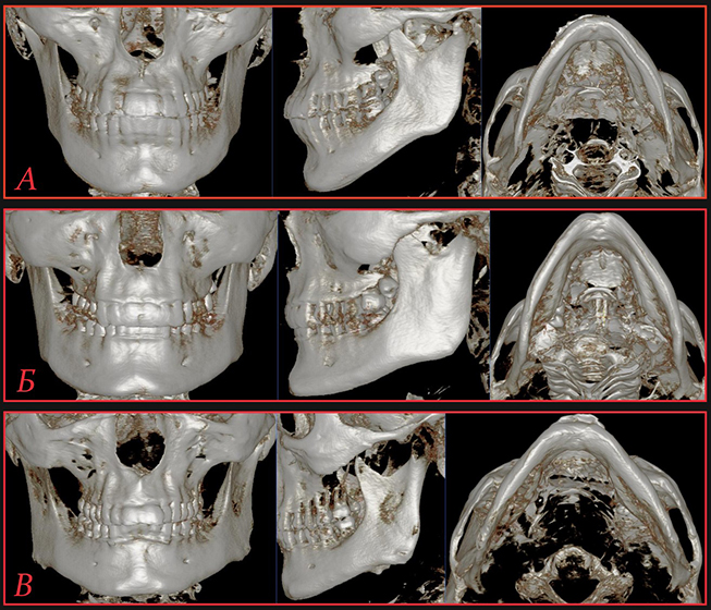
Research materials and methods: the work used dry bone preparations of whole or fragmented human skulls of both sexes in the amount of 39 units, from the collection of the Department of Human Anatomy of the Kharkiv National Medical University, and the results of CT studies of the head of adult people without existing bone tissue pathologies, totaling 85 observations. The basis of establishing a craniotype is the principle of calculating the general facial or facial index, which allows classifying anatomical objects according to the shape of the head structure.
Research results: the straight length of the mandible in adult leptoprosops males is from 88.5 mm to 102.4 mm, for women - from 86.3 mm to 100.7 mm; in mesoprosops men, this parameter gradually decreases to the level - from 81.3 mm to 95.7 mm, in women - from 80.7 mm to 94.9 mm; in euryprosops, the index is the smallest and ranges from 79.7 mm to 91.5 mm in males and from 78.5 mm to 90.8 mm in females. The opposite trend with significant ranges of variation is established for the angular width of the bone. Thus, in male leptoprosops, this parameter was determined from 84.6 mm to 97.5 mm, in female representatives - from 83.6 mm to 96.3 mm; in mesoprosops men, it increased from 89.1 mm to 105.3 mm, in women - from 87.9 mm to 103.1 mm; in euryprosops, regardless of sex, it reached its peak values, from 94.5 mm to 116.1 mm and from 92.7 mm to 114.1 mm, respectively. The height of the mandibular body also showed a certain dependence on the type of skull structure, in leptoprosops men it tended to the highest values and was fixed at the level from 29.1 mm to 38.9 mm, as well as in women - from 27.5 mm to 37.8 mm; at the same time, in mesoprosops men, the size decreased from 25.9 mm to 36.3 mm, in women, in turn, from 24.6 mm to 35.1 mm; in euryprosops men, this parameter ranged from 22.3 mm to 33.1 mm, and next to women - from 21.9 mm to 31.9 mm, it was at the level of the lowest indicators. When analyzing such a complex and multidirectional parameter as the arch of the mandible, it was also possible to obtain a characteristic of its dependence on the type of structure of the facial department of the skull. It was established that the range with the smallest values of the length of the arc is characteristic of leptoprosops, ranging from 135.8 mm to 149.4 mm in males and from 133.5 mm to 147.3 mm in females; average indices are characteristic of men - from 139.1 mm to 154.6 mm and women - from 136.4 mm to 151.2 mm with a mesoprosopic craniotype; in euryprosops of both sexes, the index tends to the greatest values at the level of 141.2 mm to 158.3 mm and from 139.7 mm to 155.7 mm, respectively.
Conclusions: the main craniometric parameters of the mandible are significantly dependent on the type of structure of the facial department of the skull. Thus, the leptoprosopic craniotype is characterized by maximum values of longitudinal and height dimensions with minimal indicators of width and arc length. Mesoprosops are characterized by the definition of intermediate, averaged values equidistant from marginal, terminal forms. In representatives with the euryprosopic type of skull structure, a significant decrease in the length and height of the jaw with a significant increase, up to the maximum values, width and length of the arch was observed. At the same time, unlike leptoprosops, which had a shortened and pointed arch, in euryprosops, the arch tended to be smoothed and lengthened. The assessment by sex leads to the fact that all sizes of the lower jaw predominate in men, but, nevertheless, a certain number of differences in indicators were within the limits of statistical error, which does not allow making absolute conclusions in this matter
References
- Holovatskyi, A. S., Cherkasov, V. H., Sapin, M. R., Parakhin, A. I., Kovalchuk, O. I. (2013). Anatomiia liudyny. Vinnytsia: Nova knyha.
- Sazonova, O. M., Vovk, O. Yu., Vovk, Yu. M., Hordiichuk, D. O., Dubina, S. O. (2018). Craniometric characteristic of the visceral skull in adulthood. Biomedical and Biosocial Anthropology, 32, 5–12. https://doi.org/10.31393/bba32-2018-01
- Sazonova, O., Vovk, O., Hordiichuk, D., Ikramov, V., Onashko, Y. (2017). Establishing the range of variability of the skull structures in adulthood. Journal of Education, Health and Sport, 7 (12), 656–664. http://dx.doi.org/10.5281/zenodo.1478808
- Li, K., Chow, W., Zhu, Z., Tai, Y., Song, J., Liu, Y., Luo, E. (2023). Comparison of Effects between Total Maxillary Setback Osteotomy and Anterior Maxillary Segmental Osteotomy on Nasolabial Morphology. Plastic & Reconstructive Surgery, 152 (6), 1076e–1087e. https://doi.org/10.1097/prs.0000000000010447
- Depeyre, A., Touzet-Roumazeille, S., Lauwers, L., Raoul, G., Ferri, J. (2016). Retrospective evaluation of 211 patients with maxillofacial reconstruction using parietal bone graft for implants insertion. Journal of Cranio-Maxillofacial Surgery, 44 (9), 1162–1169. https://doi.org/10.1016/j.jcms.2016.06.034
- Vovk, Yu. N., Vovk, O. Yu., Ikramov, V. B., Shmargalev, A. A., Malahov, S. S. (2016). Practical value of the individual anatomical variability for modern craniology. Clinical anatomy and operative surgery, 15 (1), 105–109.
- Vovk, Yu. M., Vovk, O. Yu. (2019). Indyvidualna anatomichna minlyvist ta yii kliniko-morfolohichne znachennia. Kharkiv: FOP Brovin O.V., 187.
- Celebi, A. A., Kau, C. H., Femiano, F., Bucci, L., Perillo, L. (2018). A Three-Dimensional Anthropometric Evaluation of Facial Morphology. Journal of Craniofacial Surgery, 29 (2), 304–308. https://doi.org/10.1097/scs.0000000000004110
- Verner, F. S., Roque-Torres, G. D., Ramírez-Sotello, L. R., Devito, K. L., Almeida, S. M. (2017). Analysis of the correlation between dental arch and articular eminence morphology: a cone beam computed tomography study. Oral Surgery, Oral Medicine, Oral Pathology and Oral Radiology, 124 (4), 420–431. https://doi.org/10.1016/j.oooo.2017.07.004
- Sazonova, O. M. (2019). Individual Anatomical Variability of Mandibular Alveolar Arc in Adulthood. Ukrainian Journal of Medicine, Biology and Sport, 4 (2), 87–93. https://doi.org/10.26693/jmbs04.02.087
- Sazonova, O. M., Vovk, O. Yu., Hordiichuk, D. O., Dubina, S. O. (2019). Osteometric mandibular characteristics with considering craniotype. Bulletin of Problems Biology and Medicine, 1 (1), 299–303. https://doi.org/10.29254/2077-4214-2019-1-1-148-299-303
Downloads
Published
How to Cite
Issue
Section
License
Copyright (c) 2024 Ruslan Yakymenko, Oleh Vovk

This work is licensed under a Creative Commons Attribution 4.0 International License.
Our journal abides by the Creative Commons CC BY copyright rights and permissions for open access journals.
Authors, who are published in this journal, agree to the following conditions:
1. The authors reserve the right to authorship of the work and pass the first publication right of this work to the journal under the terms of a Creative Commons CC BY, which allows others to freely distribute the published research with the obligatory reference to the authors of the original work and the first publication of the work in this journal.
2. The authors have the right to conclude separate supplement agreements that relate to non-exclusive work distribution in the form in which it has been published by the journal (for example, to upload the work to the online storage of the journal or publish it as part of a monograph), provided that the reference to the first publication of the work in this journal is included.