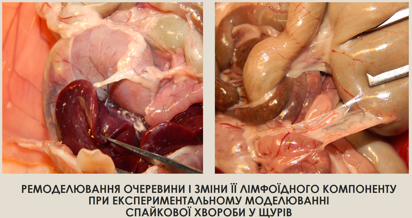
Ремоделювання очеревини і зміни її лімфоїдного компоненту при експериментальному моделюванні спайкової хвороби у щурів
DOI:
https://doi.org/10.15587/2519-8025.2024.301278Ключові слова:
очеревина, шлунково-кишковий тракт, лімфоцити, імунітет, гістологічні зміни, щури, морфометрія, мікроскопія, імунна система, спайкова хворобаАнотація
Вікові зміни, запальні процеси, хірургічні втручання та гетерогенні патологічні впливи на фізіологічні процеси очеревини призводять до певних змін у структурі компонентів очеревини, що призводить до ремоделювання тканинних структур черевної порожнини. За даними літератури, найчастішим наслідком подібного ремоделювання тканинних структур очеревини є розвиток спайкового процесу. На сьогоднішній день відсутні дані щодо вивчення лімфоїдного компонента тканин брижі, що і постає метою подальших досліджень.
Мета роботи: вивчити процес ремоделювання очеревинної тканини при експериментальному спайкоутворенні та специфіку змін її лімфоїдного компонента у щурів порівняно з нормою.
Матеріали і методи дослідження: препарування, макроскопічний, мікроскопічний, гістологічний (виготовлення плівкових препаратів), забарвлення препаратів гематоксиліном та еозином, математичний (морфометричні сітки – підрахунок кількості імунокомпетентних клітин на 1000 мкм2 стандартної площі), статистична обробка за Стьюдентом.
Результати: експериментальна спайкова хвороба характеризується поступовим процесом ремоделюванням тканин брижі тонкої кишки і, як наслідок, утворенням сполучнотканинних новоутворень. Брижа тонкої кишки втрачає еластичність і рухливість та значно потовщується. Процес формування експериментальної адгезії супроводжувався динамічними змінами кількості лімфоцитів.
Висновки: дані структури є тонкими і гомогенними на 7-й день; твердими, щільними і зернистими на 14-й день; містять тверді конгломерати гетерогенних структур на 21-й день після ін'єкції тальку. Кількість лімфоцитів у цій структурі поступово зростає: на 7 добу - на 2 % у тварин ІІ групи, на 14 добу - на 30 % у тварин ІІІ групи та на 21 добу - на 36 % у тварин IV групи, порівняно з тваринами інтактної групи
Посилання
- Lichtenstein, G. R., Loftus, E. V., Isaacs, K. L., Regueiro, M. D., Gerson, L. B., Sands, B. E. (2018). ACG Clinical Guideline: Management of Crohn’s Disease in Adults. American Journal of Gastroenterology, 113 (4), 481–517. https://doi.org/10.1038/ajg.2018.27
- Eskildsen, M. P. R., Kalliokoski, O., Boennelycke, M., Lundquist, R., Settnes, A., Løkkegaard, E. (2022). Autologous Blood-Derived Patches Used as Anti-adhesives in a Rat Uterine Horn Damage Model. Journal of Surgical Research, 275, 225–234. https://doi.org/10.1016/j.jss.2022.02.008
- Hryn, V. H. (2018). General anatomical characteristics of small intestine in white rats. Actual Problems of the Modern Medicine: Bulletin of Ukrainian Medical Stomatological Academy, 18 (4), 88–93. https://doi.org/10.31718/2077-1096.18.4.88
- Krishnan, V., Tallapragada, S., Schaar, B., Kamat, K., Chanana, A. M., Zhang, Y. et al. (2020). Omental macrophages secrete chemokine ligands that promote ovarian cancer colonization of the omentum via CCR1. Communications Biology, 3 (1), 524–529. https://doi.org/10.1038/s42003-020-01246-z
- Bukata, V. V. (2017). Experimental Research Of Efficient Use Of Barrier Methods For Preventing Adhesions In The Abdominal Cavity. Hospital Surgery. Journal Named by L. Ya. Kovalchuk, 1, 58–64. https://doi.org/10.11603/2414-4533.2017.1.7337
- Ksonz, I. V. (2015). Clinical effectiveness of anti-adhesive drugs in treatment and prevention of adhesive intestinal obstruction in children. Aktualni problemy suchasnoi medytsyny: Visnyk ukrainskoi medychnoi stomatolohichnoi akademii, 15 (3), 125–197.
- Ksyonz, I. V., Kostylenko, Y., Liakhovskyi, V. I., Konoplitskyi, V. S., Maksimovskyi, V. Y. (2023). Milky spots in the greater omentum. Actual Problems of the Modern Medicine: Bulletin of Ukrainian Medical Stomatological Academy, 23 (2.2), 135–140. https://doi.org/10.31718/2077-1096.23.2.2.135
- Marushko, Y., Hyshchak, T., Chabanovich, O. (2021). The Main Mechanisms of the Effect of Intestinal Microflora on the Immune System and Their Importance in Clinical Practice. Family Medicine, 4, 19–27. https://doi.org/10.30841/2307-5112.4.2021.249409
- Yushkov, B., Sarapultsev, A., Sarapultsev, G. (2020). Major Characteristics of Experimental Models of Abdominal Adhesions. Journal of Experimental and Clinical Surgery, 13 (2), 157–162. https://doi.org/10.18499/2070-478x-2020-13-2-157-162
- Paidarkina, A. (2023). Problema vyboru eksperymentalnoi modeli spaikovoi khvoroby. Moloda Nauka-2023. Zaporizhzhia, 257–259.
- Paidarkina, A., Kushch, O. (2023) Study of the morphological features of the peritoneum of white rats and the method of its extraction. Morphologia, 17 (3), 163–167.
- Terri, M., Trionfetti, F., Montaldo, C., Cordani, M., Tripodi, M., Lopez-Cabrera, M., Strippoli, R. (2021). Mechanisms of Peritoneal Fibrosis: Focus on Immune Cells–Peritoneal Stroma Interactions. Frontiers in Immunology, 12. https://doi.org/10.3389/fimmu.2021.607204
- Melnichenko, M. G., Kvashnina, A. A. (2019). Peritoneal regeneration and pathogenesis of postoperative peritoneal adhesions formation. Surgery of Ukraine, 3, 88–93. https://doi.org/10.30978/su2019-3-88
- Murando, F., Peloso, A., & Cobianchi, L. (2019). Experimental Abdominal Sepsis: Sticking to an Awkward but Still Useful Translational Model. Mediators of Inflammation, 2019, 1–8. https://doi.org/10.1155/2019/8971036
- Stepanchuk, A. P., Fedorchenko, I. L., Tarasenko, Ya. A., Tykhonova, O. O., Filenko, B. M. (2021). Histostructure of the Normal Human Greater Omentum and in Peritonitis. Ukrainian Journal of Medicine, Biology and Sports, 6 (5), 127–133. https://doi.org/10.26693/jmbs06.05.127
- Hu, Q., Xia, X., Kang, X., Song, P., Liu, Z., Wang, M., Guan, W., Liu, S. (2021). A review of physiological and cellular mechanisms underlying fibrotic postoperative adhesion. International Journal of Biological Sciences, 17 (1), 298–306. https://doi.org/10.7150/ijbs.54403
- Khashchuk, V. S. (2021) Mechanisms of adhesive process development in peritoneal cavity (literature review). Klinichna ta eksperymentalʹna patolohiya, 20 (4 (78)), 137–145.
- Isaza-Restrepo, A., Martin-Saavedra, J. S., Velez-Leal, J. L., Vargas-Barato, F., Riveros-Dueñas, R. (2018). The Peritoneum: Beyond the Tissue – A Review. Frontiers in Physiology, 9, 738–743. https://doi.org/10.3389/fphys.2018.00738
- Cleypool, C. G. J., Schurink, B., van der Horst, D. E. M., Bleys, R. L. A. W. (2019). Sympathetic nerve tissue in milky spots of the human greater omentum. Journal of Anatomy, 236 (1), 156–164. https://doi.org/10.1111/joa.13077
- Schurink, B., Cleypool, C. G. J., Bleys, R. L. A. W. (2019). A rapid and simple method for visualizing milky spots in large fixed tissue samples of the human greater omentum. Biotechnic & Histochemistry, 94 (6), 429–434. https://doi.org/10.1080/10520295.2019.1583375
- Maksymenko, O. S., Hryn, V. H. (2023). The Greater Omentum of White Rats: Structural and Functional Characteristics and its Role in Peritonitis. Ukrainian Journal of Medicine, Biology and Sport, 8 (1), 22–29. https://doi.org/10.26693/jmbs08.01.022

##submission.downloads##
Опубліковано
Як цитувати
Номер
Розділ
Ліцензія
Авторське право (c) 2024 Anastasia Paidarkina, Oksana Kushch

Ця робота ліцензується відповідно до Creative Commons Attribution 4.0 International License.
Наше видання використовує положення про авторські права Creative Commons CC BY для журналів відкритого доступу.
Автори, які публікуються у цьому журналі, погоджуються з наступними умовами:
1. Автори залишають за собою право на авторство своєї роботи та передають журналу право першої публікації цієї роботи на умовах ліцензії Creative Commons CC BY, котра дозволяє іншим особам вільно розповсюджувати опубліковану роботу з обов'язковим посиланням на авторів оригінальної роботи та першу публікацію роботи у цьому журналі.
2. Автори мають право укладати самостійні додаткові угоди щодо неексклюзивного розповсюдження роботи у тому вигляді, в якому вона була опублікована цим журналом (наприклад, розміщувати роботу в електронному сховищі установи або публікувати у складі монографії), за умови збереження посилання на першу публікацію роботи у цьому журналі.


