Improving the accuracy of perfusion map generation using dynamic susceptibility contrast magnetic resonance imaging data based on recurrent neural networks
DOI:
https://doi.org/10.15587/1729-4061.2026.360606Keywords:
perfusion parameters, perfusion with dynamic susceptibility contrast, magnetic resonance imagingAbstract
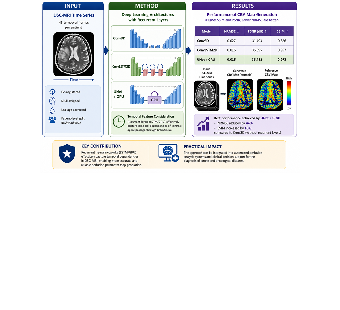
This study explores the process of generating perfusion parameter maps from time series of DSC-MRI images of the brain. The task addressed relates to the inaccuracy and inefficiency of perfusion map generation due to insufficient analysis of temporal features, dependence on the arterial inflow function, as well as excessive computational complexity of the models.
This paper proposes an approach to generating perfusion parameter maps using neural networks that directly process time series of perfusion images. Three deep learning architectures have been designed and experimentally investigated, differing in the way in which temporal information is taken into account and the presence of recurrent layers. The evaluation was performed on an open medical data set with patient-level sample distribution to prevent information leakage between the training and test parts.
A controlled comparison of models with and without recurrent layers was performed to determine the impact of explicitly taking into account temporal dependences on the accuracy of map generation. The normalized root mean square error decreased from 0.027 to 0.016 and 0.015. The structural similarity index increased from 0.826 to 0.957 and 0.973. The peak signal-to-noise ratio increased from 31.493 to 36.095 and 36.412.
Additional comparison to approaches reported in other studies showed that the proposed architecture with recurrent layers demonstrates competitive or higher values of image quality metrics. The results confirm the feasibility of using neural networks with recurrent layers for more accurate generation of perfusion parameter maps.
The practical significance is the possibility of integrating the approach into automated perfusion analysis systems and clinical decision support in the diagnosis of stroke and brain tumors
References
- Feigin, V. L., Stark, B. A., Johnson, C. O., Roth, G. A., Bisignano, C., Abady, G. G. et al. (2021). Global, regional, and national burden of stroke and its risk factors, 1990–2019: a systematic analysis for the Global Burden of Disease Study 2019. The Lancet Neurology, 20 (10), 795–820. https://doi.org/10.1016/s1474-4422(21)00252-0
- O’Connor, J. P. B., Tofts, P. S., Miles, K. A., Parkes, L. M., Thompson, G., Jackson, A. (2011). Dynamic contrast-enhanced imaging techniques: CT and MRI. The British Journal of Radiology, 84, S112–S120. https://doi.org/10.1259/bjr/55166688
- Alkhimova, S., Sorokina, V., Kabala, I. (2026). Comparative assessment of commonly used color lookup tables to determine key performance indicators for perfusion map data visualization. Technology Audit and Production Reserves, 1 (2 (87)), 85–92. https://doi.org/10.15587/2706-5448.2026.352787
- Diumin, O. D., Alkhimova, S. M. (2024). Generation of perfusion map images using deep learning methods. Nauka i tekhnika sohodni, 11 (39), 849–866. https://doi.org/10.52058/2786-6025-2024-10(38)-849-866
- Alkhimova, S. M., Diumin, O. D. (2024). Issues of automated perfusion analysis based on dynamic susceptibility contrast MRI. Nauka i tekhnika sohodni, 8 (36), 859–875. https://doi.org/10.52058/2786-6025-2024-8(36)-859-875
- Østergaard, L., Weisskoff, R. M., Chesler, D. A., Gyldensted, C., Rosen, B. R. (1996). High resolution measurement of cerebral blood flow using intravascular tracer bolus passages. Part I: Mathematical approach and statistical analysis. Magnetic Resonance in Medicine, 36 (5), 715–725. https://doi.org/10.1002/mrm.1910360510
- Wu, O., Østergaard, L., Weisskoff, R. M., Benner, T., Rosen, B. R., Sorensen, A. G. (2003). Tracer arrival timing‐insensitive technique for estimating flow in MR perfusion‐weighted imaging using singular value decomposition with a block‐circulant deconvolution matrix. Magnetic Resonance in Medicine, 50 (1), 164–174. https://doi.org/10.1002/mrm.10522
- Kudo, K., Christensen, S., Sasaki, M., Straka, M., Fujiwara, S., Ishizaka, K. et al. (2010). Acuracy and reliability of post-processing software for DSC MR perfusion: Quantitative analysis by digital phantom data. In Proceedings of the 18th Annual Meetings of the ISMRM. Available at: https://archive.ismrm.org/2010/1791.html
- Gautam, R., Sivaswamy, J., Varma, R. (2012). An efficient, bolus-stage based method for motion correction in perfusion weighted MRI. In Proceedings of the 21st International Conference on Pattern Recognition (ICPR2012), 145–148. IEEE. Available at: https://ieeexplore.ieee.org/abstract/document/6460093
- Alkhimova, S. (2018). Detection of perfusion ROI as a quality control in perfusion analysis. In Science, research, development. arXiv. https://doi.org/10.48550/arXiv.1902.01855
- Alkhimova, S. (2019). Impact of perfusion ROI detection to the quality of CBV perfusion map. Technology Audit and Production Reserves, 5 (2 (49)), 27–30. https://doi.org/10.15587/2312-8372.2019.182789
- Alkhimova, S. M., Sliusar, S. V. (2019). Analysis of effectiveness of thresholding in perfusion roi detection on T2-weighted MR images with abnormal brain anatomy. KPI Science News, 4, 35–43. https://doi.org/10.20535/kpi-sn.2019.4.180237
- Alkhimova, S., Krenevych, A. (2019). Brain tissues segmentation on mr perfusion images using CUSUM filter for boundary pixels. International Journal of Computing, 18 (2), 127–134. https://doi.org/10.47839/ijc.18.2.1411
- Alkhimova, S. (2019). CUSUM Filter for Brain Segmentation on DSC Perfusion MR Head Scans with Abnormal Brain Anatomy. Proceedings of the 2019 International Conference on Intelligent Medicine and Image Processing, 43–47. https://doi.org/10.1145/3332340.3332357
- Alkhimova, S., Diumin, O. (2023). Deep learning-based neural network for regions of interest retrieval in T2*-weighted brain perfusion MRI. Nauka I Tekhnika Sohodni, 14 (14). https://doi.org/10.52058/2786-6025-2022-14(14)-301-314
- Fieselmann, A., Kowarschik, M., Ganguly, A., Hornegger, J., Fahrig, R. (2011). Deconvolution-Based CT and MR Brain Perfusion Measurement: Theoretical Model Revisited and Practical Implementation Details. International Journal of Biomedical Imaging, 2011, 467563. https://doi.org/10.1155/2011/467563
- Nastenko, I., Linnik, M., Honcharuk, M., Davydovych, I., Lutchenko, V., Babenko, V., Dolinchuk, L. (2025). Results of Machine Learning Applications in the Study of COVID-19 Associated Cardiopulmonary Pathology Using Computed Tomography Data. Innovative Biosystems and Bioengineering, 9 (4), 16–27. https://doi.org/10.20535/ibb.2025.9.4.325335
- Sethi, S., Reddy, S., Sakarvadia, M., Serotte, J., Nwaudo, D., Maassen, N., Shi, L. (2025). Toward non-invasive diagnosis of Bankart lesions with deep learning. Medical Imaging 2025: Computer-Aided Diagnosis, 134. https://doi.org/10.1117/12.3046251
- Xie, H., Gan, W., Bayerlein, R., Zhou, B., Chen, M.-K., Kulon, M. et al. (2026). Dose-aware diffusion model for 3D PET image denoising: Multi-institutional validation with reader study and real low-dose data. Medical Image Analysis, 111, 104039. https://doi.org/10.1016/j.media.2026.104039
- Mahmood, M. F. (2024). Recognition and Categorization of Blood Groups by Machine Learning and Image Processing Method. Innovative Biosystems and Bioengineering, 8 (2), 53–68. https://doi.org/10.20535/ibb.2024.8.2.298201
- Ho, K. C., Scalzo, F., Sarma, K. V., El-Saden, S., Arnold, C. W. (2016). A temporal deep learning approach for MR perfusion parameter estimation in stroke. 2016 23rd International Conference on Pattern Recognition (ICPR), 1315–1320. https://doi.org/10.1109/icpr.2016.7899819
- Hess, A., Meier, R., Kaesmacher, J., Jung, S., Scalzo, F., Liebeskind, D. et al. (2019). Synthetic Perfusion Maps: Imaging Perfusion Deficits in DSC-MRI with Deep Learning. Brainlesion: Glioma, Multiple Sclerosis, Stroke and Traumatic Brain Injuries, 447–455. https://doi.org/10.1007/978-3-030-11723-8_45
- Asaduddin, M., Roh, H. G., Kim, H. J., Kim, E. Y., Park, S.-H. (2022). Perfusion Maps Acquired From Dynamic Angiography MRI Using Deep Learning Approaches. Journal of Magnetic Resonance Imaging, 57 (2), 456–469. https://doi.org/10.1002/jmri.28315
- Kossen, T., Madai, V. I., Mutke, M. A., Hennemuth, A., Hildebrand, K., Behland, J. et al. (2023). Image-to-image generative adversarial networks for synthesizing perfusion parameter maps from DSC-MR images in cerebrovascular disease. Frontiers in Neurology, 13. https://doi.org/10.3389/fneur.2022.1051397
- Talebi, S., Gai, S., Sossin, A., Zhu, V., Tong, E., Mofrad, M. R. K. (2024). Deep Learning for Perfusion Cerebral Blood Flow (CBF) and Volume (CBV) Predictions and Diagnostics. Annals of Biomedical Engineering, 52 (6), 1568–1575. https://doi.org/10.1007/s10439-024-03471-7
- Cao, A., Le, P.-Y., Qie, Z., Guo, Y., Zaman, A., Lu, J. et al. (2024). Quantitative Perfusion Maps Using a Novelty Spatiotemporal Convolutional Neural Network. 2024 IEEE International Symposium on Biomedical Imaging (ISBI), 1–5. https://doi.org/10.1109/isbi56570.2024.10635136
- McKinley, R., Hung, F., Wiest, R., Liebeskind, D. S., Scalzo, F. (2018). A Machine Learning Approach to Perfusion Imaging With Dynamic Susceptibility Contrast MR. Frontiers in Neurology, 9. https://doi.org/10.3389/fneur.2018.00717
- Ahmed, Md. M., Hossain, Md. M., Islam, Md. R., Ali, Md. S., Nafi, A. A. N., Ahmed, Md. F. et al. (2024). Brain tumor detection and classification in MRI using hybrid ViT and GRU model with explainable AI in Southern Bangladesh. Scientific Reports, 14 (1). https://doi.org/10.1038/s41598-024-71893-3
- Murase, K., Nakamoto, A., Tomiyama, N. (2024). Performance of recurrent neural networks with Monte Carlo dropout for predicting pharmacokinetic parameters from dynamic contrast‐enhanced magnetic resonance imaging data. Journal of Applied Clinical Medical Physics, 26 (2). https://doi.org/10.1002/acm2.14586
- Bakas, S., Sako, C., Akbari, H., Bilello, M., Sotiras, A., Shukla, G. et al. (2022). The University of Pennsylvania glioblastoma (UPenn-GBM) cohort: advanced MRI, clinical, genomics, & radiomics. Scientific Data, 9 (1). https://doi.org/10.1038/s41597-022-01560-7
Downloads
Published
How to Cite
Issue
Section
License
Copyright (c) 2026 Oleksii Diumin, Svitlana Alkhimova

This work is licensed under a Creative Commons Attribution 4.0 International License.
The consolidation and conditions for the transfer of copyright (identification of authorship) is carried out in the License Agreement. In particular, the authors reserve the right to the authorship of their manuscript and transfer the first publication of this work to the journal under the terms of the Creative Commons CC BY license. At the same time, they have the right to conclude on their own additional agreements concerning the non-exclusive distribution of the work in the form in which it was published by this journal, but provided that the link to the first publication of the article in this journal is preserved.
A license agreement is a document in which the author warrants that he/she owns all copyright for the work (manuscript, article, etc.).
The authors, signing the License Agreement with TECHNOLOGY CENTER PC, have all rights to the further use of their work, provided that they link to our edition in which the work was published.
According to the terms of the License Agreement, the Publisher TECHNOLOGY CENTER PC does not take away your copyrights and receives permission from the authors to use and dissemination of the publication through the world's scientific resources (own electronic resources, scientometric databases, repositories, libraries, etc.).
In the absence of a signed License Agreement or in the absence of this agreement of identifiers allowing to identify the identity of the author, the editors have no right to work with the manuscript.
It is important to remember that there is another type of agreement between authors and publishers – when copyright is transferred from the authors to the publisher. In this case, the authors lose ownership of their work and may not use it in any way.