Devising a comprehensive approach to diagnosing breast cancer subtypes automatically based on deep neural networks
DOI:
https://doi.org/10.15587/1729-4061.2025.344041Keywords:
Attention U-Net, genetic algorithm, neural network architecture optimization, IHC images, biomedical image segmentation, automatic diagnosing of breast cancerAbstract
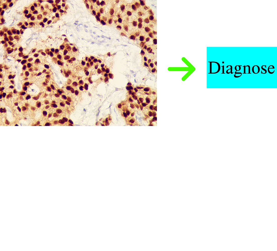
This study investigates the process of analyzing immunohistochemical images of breast cancer. The study has contributed to solving the task of a standardized and objective approach to the quantitative assessment of immunohistochemical biomarkers, which would minimize inter-individual variability in assessments and could be computationally efficient for the analysis of biomedical images.
This paper aims to balance model complexity and generalization by using evolutionary algorithms to tune deep neural networks for biomedical tasks, analyzing how network structure affects performance.
Experiments were conducted on the segmentation of immunohistochemical images on 13 different architectures of neural networks. The evaluation was performed using five accuracy metrics, which allowed for an objective comparison of model performance. The use of a genetic algorithm to optimize the neural network architecture made it possible to adaptively find combinations of parameters, in particular the number of layers and the size of the base filter. The evolutionary approach enabled effective exploration of configuration space, which led to an increase in the Dice metric to 0.74. The resulting increase in accuracy indicates the model’s improved ability to segment images with different characteristics, demonstrating the practical effectiveness of the proposed approach for biomedical diagnosis tasks.
The optimized architecture was used to design a system for diagnosing breast cancer automatically based on neural networks, in particular for the method of automatic diagnosis of breast cancer subtypes. That contributed to improving the accuracy of biomedical image analysis, which could help improve the diagnostic process in clinical practice
References
- Hamet, P., Tremblay, J. (2017). Artificial intelligence in medicine. Metabolism, 69, S36–S40. https://doi.org/10.1016/j.metabol.2017.01.011
- Briganti, G., Le Moine, O. (2020). Artificial Intelligence in Medicine: Today and Tomorrow. Frontiers in Medicine, 7. https://doi.org/10.3389/fmed.2020.00027
- Litjens, G., Kooi, T., Bejnordi, B. E., Setio, A. A. A., Ciompi, F., Ghafoorian, M. et al. (2017). A survey on deep learning in medical image analysis. Medical Image Analysis, 42, 60–88. https://doi.org/10.1016/j.media.2017.07.005
- Srikantamurthy, M. M., Rallabandi, V. P. S., Dudekula, D. B., Natarajan, S., Park, J. (2023). Classification of benign and malignant subtypes of breast cancer histopathology imaging using hybrid CNN-LSTM based transfer learning. BMC Medical Imaging, 23 (1). https://doi.org/10.1186/s12880-023-00964-0
- Al-Jabbar, M., Alshahrani, M., Senan, E. M., Ahmed, I. A. (2023). Analyzing Histological Images Using Hybrid Techniques for Early Detection of Multi-Class Breast Cancer Based on Fusion Features of CNN and Handcrafted. Diagnostics, 13 (10), 1753. https://doi.org/10.3390/diagnostics13101753
- Miranda Ruiz, F., Lahrmann, B., Bartels, L., Krauthoff, A., Keil, A., Härtel, S. et al. (2023). CNN stability training improves robustness to scanner and IHC-based image variability for epithelium segmentation in cervical histology. Frontiers in Medicine, 10. https://doi.org/10.3389/fmed.2023.1173616
- Zaha, D. C. (2014). Significance of immunohistochemistry in breast cancer. World Journal of Clinical Oncology, 5 (3), 382. https://doi.org/10.5306/wjco.v5.i3.382
- Aswathy M. A., Mohan, J. (2020). Analysis of Machine Learning Algorithms for Breast Cancer Detection. Handbook of Research on Applications and Implementations of Machine Learning Techniques, 1–20. https://doi.org/10.4018/978-1-5225-9902-9.ch001
- Nabok, A. I. (2023). Prevalence and incidence of breast cancer in Ukraine. Wiadomości Lekarskie, 76 (10), 2219–2223. Available at: https://www.researchgate.net/profile/Serhii-Tertyshnyi/publication/375025887_WL_Layout_10_2023/links/653bdaf73cc79d48c5b14c25/WL-Layout-10-2023.pdf#page=93
- Siegel, R. L., Kratzer, T. B., Giaquinto, A. N., Sung, H., Jemal, A. (2025). Cancer statistics, 2025. CA: A Cancer Journal for Clinicians, 75 (1), 10–45. https://doi.org/10.3322/caac.21871
- Ronneberger, O., Fischer, P., Brox, T. (2015). U-Net: Convolutional Networks for Biomedical Image Segmentation. Medical Image Computing and Computer-Assisted Intervention – MICCAI 2015, 234–241. https://doi.org/10.1007/978-3-319-24574-4_28
- Polley, M.-Y. C., Leung, S. C. Y., McShane, L. M., Gao, D., Hugh, J. C., Mastropasqua, M. G. et al. (2013). An International Ki67 Reproducibility Study. JNCI: Journal of the National Cancer Institute, 105 (24), 1897–1906. https://doi.org/10.1093/jnci/djt306
- Kumar, N., Gupta, R., Gupta, S. (2020). Whole Slide Imaging (WSI) in Pathology: Current Perspectives and Future Directions. Journal of Digital Imaging, 33 (4), 1034–1040. https://doi.org/10.1007/s10278-020-00351-z
- Siddique, N., Paheding, S., Elkin, C. P., Devabhaktuni, V. (2021). U-Net and Its Variants for Medical Image Segmentation: A Review of Theory and Applications. IEEE Access, 9, 82031–82057. https://doi.org/10.1109/access.2021.3086020
- Mehta, R., Arbel, T. (2019). 3D U-Net for Brain Tumour Segmentation. Brainlesion: Glioma, Multiple Sclerosis, Stroke and Traumatic Brain Injuries, 254–266. https://doi.org/10.1007/978-3-030-11726-9_23
- Chen, W., Liu, B., Peng, S., Sun, J., Qiao, X. (2019). S3D-UNet: Separable 3D U-Net for Brain Tumor Segmentation. Brainlesion: Glioma, Multiple Sclerosis, Stroke and Traumatic Brain Injuries, 358–368. https://doi.org/10.1007/978-3-030-11726-9_32
- Benny, S., Varma, S. L. (2021). Semantic Segmentation in Immunohistochemistry Breast Cancer Image using Deep Learning. 2021 International Conference on Advances in Computing, Communication, and Control (ICAC3), 1–3. https://doi.org/10.1109/icac353642.2021.9697264
- Benny, S., Varma, S. L. (2023). Attention-enhanced residual U-Net for nucleus segmentation in immunohistochemistry images. International Journal of Applied Engineering & Technology, 5 (4), 1266–1283. Available at: https://romanpub.com/resources/ijaet20v5-4-2023-138.pdf
- Mahanta, L. B., Hussain, E., Das, N., Kakoti, L., Chowdhury, M. (2021). IHC-Net: A fully convolutional neural network for automated nuclear segmentation and ensemble classification for Allred scoring in breast pathology. Applied Soft Computing, 103, 107136. https://doi.org/10.1016/j.asoc.2021.107136
- Kromp, F., Fischer, L., Bozsaky, E., Ambros, I. M., Dorr, W., Beiske, K. et al. (2021). Evaluation of Deep Learning Architectures for Complex Immunofluorescence Nuclear Image Segmentation. IEEE Transactions on Medical Imaging, 40 (7), 1934–1949. https://doi.org/10.1109/tmi.2021.3069558
- Xu, S., Li, G., Song, H., Wang, J., Wang, Y., Li, Q. (2024). GeNSeg-Net: A General Segmentation Framework for Any Nucleus in Immunohistochemistry Images. Proceedings of the 32nd ACM International Conference on Multimedia, 4475–4484. https://doi.org/10.1145/3664647.3681441
- Aboudessouki, A., Ali, Kh. M., Elsharkawy, M., Alksas, A., Mahmoud, A., Khalifa, F. et al. (2023). Automated Diagnosis of Breast Cancer Using Deep Learning-Based Whole Slide Image Analysis of Molecular Biomarkers. 2023 IEEE International Conference on Image Processing (ICIP). https://doi.org/10.1109/icip49359.2023.10222479
- Tkachova, O. V., Melnyk, H. M., Pitsun, O. Y., Datsko, T. V., Klishch, I. M., Derysh, B. B. (2023). A. s. No. 118979. Baza danykh tsyfrovykh imunohistokhimichnykh zobrazhen raku molochnoi zalozy «IHCDBI». declareted: 10.05.2023; published: 31.07.2023, Bul. No. 76.
- Huynh, N. (2023). Understanding evaluation metrics in Medical Image Segmentation. Available at: https://medium.com/mastering-data-science/understanding-evaluation-metrics-in-medical-image-segmentation-d289a373a3f
- WBRT pislia BCS. Rak molochnoi zalozy na rannikh stadiyakh: Klinichna nastanova, zasnovana na dokazakh (2024). Ministerstvo okhorony zdorovia Ukrainy, 27–28. Available at: https://www.dec.gov.ua/wp-content/uploads/2025/02/kn_2025_rannij-rmz.pdf
- Berezsky, O., Pitsun, O., Melnyk, G., Datsko, T., Izonin, I., Derysh, B. (2023). An Approach toward Automatic Specifics Diagnosis of Breast Cancer Based on an Immunohistochemical Image. Journal of Imaging, 9 (1), 12. https://doi.org/10.3390/jimaging9010012
- Cardoso, F., Kyriakides, S., Ohno, S., Penault-Llorca, F., Poortmans, P., Rubio, I. T. et al. (2019). Early breast cancer: ESMO Clinical Practice Guidelines for diagnosis, treatment and follow-up. Annals of Oncology, 30 (8), 1194–1220. https://doi.org/10.1093/annonc/mdz173
- Liashchynskyi, P. B., Berezsky, O. M. (2024). Computer diagnostic systems: methods and tools. Ukrainian Journal of Information Technology, 6 (2), 57–63. https://doi.org/10.23939/ujit2024.02.057
- Berezsky, O. M., Liashchynskyi, P. B. (2024). Development of the architecture of a computer aided diagnosis system in medicine. Applied Aspects of Information Technology, 7 (4), 359–369. https://doi.org/10.15276/aait.07.2024.25
Downloads
Published
How to Cite
Issue
Section
License
Copyright (c) 2025 Oleh Berezsky, Pavlo Liashchynskyi, Petro Liashchynskyi, Petro Selskyy

This work is licensed under a Creative Commons Attribution 4.0 International License.
The consolidation and conditions for the transfer of copyright (identification of authorship) is carried out in the License Agreement. In particular, the authors reserve the right to the authorship of their manuscript and transfer the first publication of this work to the journal under the terms of the Creative Commons CC BY license. At the same time, they have the right to conclude on their own additional agreements concerning the non-exclusive distribution of the work in the form in which it was published by this journal, but provided that the link to the first publication of the article in this journal is preserved.
A license agreement is a document in which the author warrants that he/she owns all copyright for the work (manuscript, article, etc.).
The authors, signing the License Agreement with TECHNOLOGY CENTER PC, have all rights to the further use of their work, provided that they link to our edition in which the work was published.
According to the terms of the License Agreement, the Publisher TECHNOLOGY CENTER PC does not take away your copyrights and receives permission from the authors to use and dissemination of the publication through the world's scientific resources (own electronic resources, scientometric databases, repositories, libraries, etc.).
In the absence of a signed License Agreement or in the absence of this agreement of identifiers allowing to identify the identity of the author, the editors have no right to work with the manuscript.
It is important to remember that there is another type of agreement between authors and publishers – when copyright is transferred from the authors to the publisher. In this case, the authors lose ownership of their work and may not use it in any way.